2025
|
Collercandy, Nived; Vellas, Camille; Nayrac, Manon; Requena, Mary; Richarme, Thomas; Iscache, Anne-Laure; Latour, Justine; Barange, Karl; Alric, Laurent; Martin-Blondel, Guillaume; Serino, Matteo; Izopet, Jacques; Delobel, Pierre Cytotoxic CX3CR1+ Vδ1 T cells clonally expand in an interplay of CMV, microbiota, and HIV-1 persistence in people on antiretroviral therapy Article de journal Dans: PLoS Pathog, vol. 21, no. 9, p. e1013489, 2025, ISSN: 1553-7374. @article{pmid40920867,
title = {Cytotoxic CX3CR1+ Vδ1 T cells clonally expand in an interplay of CMV, microbiota, and HIV-1 persistence in people on antiretroviral therapy},
author = {Nived Collercandy and Camille Vellas and Manon Nayrac and Mary Requena and Thomas Richarme and Anne-Laure Iscache and Justine Latour and Karl Barange and Laurent Alric and Guillaume Martin-Blondel and Matteo Serino and Jacques Izopet and Pierre Delobel},
doi = {10.1371/journal.ppat.1013489},
issn = {1553-7374},
year = {2025},
date = {2025-09-01},
urldate = {2025-09-01},
journal = {PLoS Pathog},
volume = {21},
number = {9},
pages = {e1013489},
abstract = {Vδ1 γδ T cells are key players in innate and adaptive immunity, particularly at mucosal interfaces such as the gut. An increase in circulating Vδ1 cells has long been observed in people with HIV-1, but remains poorly understood. We performed a comprehensive characterization of Vδ1 T cells in blood and duodenal intra-epithelial lymphocytes, obtained from endoscopic mucosal biopsies of 15 people with HIV-1 on antiretroviral therapy and 15 HIV-seronegative controls, in a substudy of the ANRS EP61 GALT study (NCT02906137). We deciphered the phenotype, functional profile, single-cell transcriptome and repertoire of Vδ1 cells and unraveled their relationships with the possible triggers involved, in particular CMV and microbiota. We also assessed whether Vδ1 T cells may play a role in controlling the HIV-1 reservoir. Vδ1 T cells were mainly terminally differentiated effectors that clonally expanded in the blood with some trafficking with the gut of people with HIV-1. Most expressed CX3CR1 and displayed a highly cytotoxic profile, but low cytokine production, supported by a transcriptomic shift towards enhanced effector lymphocytes. This expansion was associated with CMV status and markers of occult replication, but also with changes in the duodenal and blood-translocated microbiota. Cytotoxic, but not IFN-γ-producing, Vδ1 T cells were negatively associated with cell-associated HIV-1 RNA in both the blood and duodenal compartments. The increase in Vδ1 T cells observed in people with HIV-1 has multiple triggers, particularly CMV and microbiota, and may in turn contribute to the control of the HIV-1 reservoir.},
keywords = {},
pubstate = {published},
tppubtype = {article}
}
Vδ1 γδ T cells are key players in innate and adaptive immunity, particularly at mucosal interfaces such as the gut. An increase in circulating Vδ1 cells has long been observed in people with HIV-1, but remains poorly understood. We performed a comprehensive characterization of Vδ1 T cells in blood and duodenal intra-epithelial lymphocytes, obtained from endoscopic mucosal biopsies of 15 people with HIV-1 on antiretroviral therapy and 15 HIV-seronegative controls, in a substudy of the ANRS EP61 GALT study (NCT02906137). We deciphered the phenotype, functional profile, single-cell transcriptome and repertoire of Vδ1 cells and unraveled their relationships with the possible triggers involved, in particular CMV and microbiota. We also assessed whether Vδ1 T cells may play a role in controlling the HIV-1 reservoir. Vδ1 T cells were mainly terminally differentiated effectors that clonally expanded in the blood with some trafficking with the gut of people with HIV-1. Most expressed CX3CR1 and displayed a highly cytotoxic profile, but low cytokine production, supported by a transcriptomic shift towards enhanced effector lymphocytes. This expansion was associated with CMV status and markers of occult replication, but also with changes in the duodenal and blood-translocated microbiota. Cytotoxic, but not IFN-γ-producing, Vδ1 T cells were negatively associated with cell-associated HIV-1 RNA in both the blood and duodenal compartments. The increase in Vδ1 T cells observed in people with HIV-1 has multiple triggers, particularly CMV and microbiota, and may in turn contribute to the control of the HIV-1 reservoir. |
F, Martinez; C, Cotineau; C, Bories; L, Culié; S, Rodriguez; C, Pérals; S, Lachambre; V, Duplan-Eche; F, Bucciarelli; B, Pignolet; R, Liblau; L, Michel; M, Aloulou; N, Fazilleau Follicular regulatory T cells promote experimental autoimmune encephalomyelitis by supporting B cell egress from germinal centers. Article de journal Dans: Science Translational Medecine, vol. 17 , no. 813, 2025. @article{,
title = {Follicular regulatory T cells promote experimental autoimmune encephalomyelitis by supporting B cell egress from germinal centers. },
author = {Martinez F and Cotineau C and Bories C and Culié L and Rodriguez S and Pérals C and Lachambre S and Duplan-Eche V and Bucciarelli F and Pignolet B and Liblau R and Michel L and Aloulou M and Fazilleau N},
doi = {10.1126/scitranslmed.ady1268},
year = {2025},
date = {2025-08-27},
urldate = {2025-08-27},
journal = {Science Translational Medecine},
volume = {17 },
number = {813},
keywords = {},
pubstate = {published},
tppubtype = {article}
}
|
Belloy, Marcy; Schmitt, Benjamin A M; Marty, Florent H; Paut, Charlotte; Bassot, Emilie; Aïda, Amel; Alis, Marine; Zahm, Margot; Chaubet, Adeline; Garnier, Hugo; Flores-Aguilar, Thelma; Roitg, Elisa; Gutierrez-Loli, Renzo; Allart, Sophie; Ecalard, Romain; Boursereau, Raphaël; Ligat, Gaëtan; Gonzalez-Dunia, Daniel; Blanchard, Nicolas; Suberbielle, Elsa Toxoplasma gondii infection and chronic IL-1 elevation drive hippocampal DNA double-strand break signaling, leading to cognitive deficits Article de journal Dans: Nat Neurosci, 2025, ISSN: 1546-1726. @article{pmid40841478,
title = {Toxoplasma gondii infection and chronic IL-1 elevation drive hippocampal DNA double-strand break signaling, leading to cognitive deficits},
author = {Marcy Belloy and Benjamin A M Schmitt and Florent H Marty and Charlotte Paut and Emilie Bassot and Amel Aïda and Marine Alis and Margot Zahm and Adeline Chaubet and Hugo Garnier and Thelma Flores-Aguilar and Elisa Roitg and Renzo Gutierrez-Loli and Sophie Allart and Romain Ecalard and Raphaël Boursereau and Gaëtan Ligat and Daniel Gonzalez-Dunia and Nicolas Blanchard and Elsa Suberbielle},
doi = {10.1038/s41593-025-02041-x},
issn = {1546-1726},
year = {2025},
date = {2025-08-01},
urldate = {2025-08-01},
journal = {Nat Neurosci},
abstract = {Chronic inflammation, resulting from infections, is characterized by increased levels of cytokines including interleukin-1 (IL-1), but little is known about how IL-1 contributes to cognitive impairment, potentially via epigenetic processes. Here we demonstrate that mice chronically infected with the parasite Toxoplasma gondii exhibit impaired spatial memory, which is dependent on neuronal IL-1 signaling and mimicked by chronic exposure to IL-1β. Both T. gondii infection and chronic IL-1β drive H2A.X-dependent DNA double-strand break signaling in hippocampal neurons and invalidating neuronal H2A.X-dependent signaling blocks memory impairments caused by either exposure. Our results highlight the instrumental role of cytokine-induced double-strand-break-dependent signaling in spatial memory defects, which may be relevant to multiple brain diseases.},
keywords = {},
pubstate = {published},
tppubtype = {article}
}
Chronic inflammation, resulting from infections, is characterized by increased levels of cytokines including interleukin-1 (IL-1), but little is known about how IL-1 contributes to cognitive impairment, potentially via epigenetic processes. Here we demonstrate that mice chronically infected with the parasite Toxoplasma gondii exhibit impaired spatial memory, which is dependent on neuronal IL-1 signaling and mimicked by chronic exposure to IL-1β. Both T. gondii infection and chronic IL-1β drive H2A.X-dependent DNA double-strand break signaling in hippocampal neurons and invalidating neuronal H2A.X-dependent signaling blocks memory impairments caused by either exposure. Our results highlight the instrumental role of cytokine-induced double-strand-break-dependent signaling in spatial memory defects, which may be relevant to multiple brain diseases. |
2024
|
Schiavone, Marion; Dagkesamanskaya, Adilya; Vieu, Pierre-Gilles; Duperray, Maëlle; Duplan-Eche, Valérie; François, Jean Marie A flow cytometry method for quantitative measurement and molecular investigation of the adhesion of bacteria to yeast cells Article de journal Dans: Sci Rep, vol. 14, no. 1, p. 20935, 2024, ISSN: 2045-2322. @article{pmid39251857,
title = {A flow cytometry method for quantitative measurement and molecular investigation of the adhesion of bacteria to yeast cells},
author = {Marion Schiavone and Adilya Dagkesamanskaya and Pierre-Gilles Vieu and Maëlle Duperray and Valérie Duplan-Eche and Jean Marie François},
doi = {10.1038/s41598-024-72030-w},
issn = {2045-2322},
year = {2024},
date = {2024-09-01},
urldate = {2024-09-01},
journal = {Sci Rep},
volume = {14},
number = {1},
pages = {20935},
abstract = {The study of microorganism interactions is important for understanding the organization and functioning of microbial consortia. Additionally, the interaction between yeast and bacteria is of interest in the field of health and nutrition area for the development of probiotics. To investigate these microbial interactions at the cellular and molecular levels, a simple, reliable, and quantitative method is proposed. We demonstrated that flow cytometry enables the measurement of interactions at a single-cell level by detecting and counting yeast cells with bound fluorescent bacteria. Imaging flow cytometry revealed that the number of bacteria attached to yeast followed a Gaussian distribution whose maximum reached 14 bacterial cells using a clinical Escherichia coli strain E22 and the laboratory yeast strain BY4741. We found that the dynamics of adhesion resemble a Langmuir adsorption model, albeit it is a rapid and almost irreversible process. This adhesion is dependent on the mannose-specific type 1 fimbriae, as E. coli mutants lacking these appendages no longer adhere to yeast. However, this type 1 fimbriae-dependent adhesion could involve additional yeast cell wall factors, since the interaction between bacteria and yeast mutants with altered mannan content remained comparable to that of wild-type yeast. In summary, flow cytometry is an appropriate method for studying bacteria-yeast adhesion, as well as for the high-throughput screening of candidate molecules likely to promote or counteract this interaction.},
keywords = {},
pubstate = {published},
tppubtype = {article}
}
The study of microorganism interactions is important for understanding the organization and functioning of microbial consortia. Additionally, the interaction between yeast and bacteria is of interest in the field of health and nutrition area for the development of probiotics. To investigate these microbial interactions at the cellular and molecular levels, a simple, reliable, and quantitative method is proposed. We demonstrated that flow cytometry enables the measurement of interactions at a single-cell level by detecting and counting yeast cells with bound fluorescent bacteria. Imaging flow cytometry revealed that the number of bacteria attached to yeast followed a Gaussian distribution whose maximum reached 14 bacterial cells using a clinical Escherichia coli strain E22 and the laboratory yeast strain BY4741. We found that the dynamics of adhesion resemble a Langmuir adsorption model, albeit it is a rapid and almost irreversible process. This adhesion is dependent on the mannose-specific type 1 fimbriae, as E. coli mutants lacking these appendages no longer adhere to yeast. However, this type 1 fimbriae-dependent adhesion could involve additional yeast cell wall factors, since the interaction between bacteria and yeast mutants with altered mannan content remained comparable to that of wild-type yeast. In summary, flow cytometry is an appropriate method for studying bacteria-yeast adhesion, as well as for the high-throughput screening of candidate molecules likely to promote or counteract this interaction. |
Lacouture, Claire; Chaves, Beatriz; Guipouy, Delphine; Houmadi, Raïssa; Duplan-Eche, Valérie; Allart, Sophie; Destainville, Nicolas; Dupré, Loïc LFA-1 nanoclusters integrate TCR stimulation strength to tune T-cell cytotoxic activity Article de journal Dans: Nature Communications, vol. 15, no. 1, p. 407, 2024, ISSN: 2041-1723, (Number: 1
Publisher: Nature Publishing Group). @article{lacouture_lfa-1_2024,
title = {LFA-1 nanoclusters integrate TCR stimulation strength to tune T-cell cytotoxic activity},
author = {Claire Lacouture and Beatriz Chaves and Delphine Guipouy and Raïssa Houmadi and Valérie Duplan-Eche and Sophie Allart and Nicolas Destainville and Loïc Dupré},
url = {https://www.nature.com/articles/s41467-024-44688-3},
doi = {10.1038/s41467-024-44688-3},
issn = {2041-1723},
year = {2024},
date = {2024-01-01},
urldate = {2024-01-01},
journal = {Nature Communications},
volume = {15},
number = {1},
pages = {407},
abstract = {T-cell cytotoxic function relies on the cooperation between the highly specific but poorly adhesive T-cell receptor (TCR) and the integrin LFA-1. How LFA-1-mediated adhesion may scale with TCR stimulation strength is ill-defined. Here, we show that LFA-1 conformation activation scales with TCR stimulation to calibrate human T-cell cytotoxicity. Super-resolution microscopy analysis reveals that >1000 LFA-1 nanoclusters provide a discretized platform at the immunological synapse to translate TCR engagement and density of the LFA-1 ligand ICAM-1 into graded adhesion. Indeed, the number of high-affinity conformation LFA-1 nanoclusters increases as a function of TCR triggering strength. Blockade of LFA-1 conformational activation impairs adhesion to target cells and killing. However, it occurs at a lower TCR stimulation threshold than lytic granule exocytosis implying that it licenses, rather than directly controls, the killing decision. We conclude that the organization of LFA-1 into nanoclusters provides a calibrated system to adjust T-cell killing to the antigen stimulation strength.},
note = {Number: 1
Publisher: Nature Publishing Group},
keywords = {},
pubstate = {published},
tppubtype = {article}
}
T-cell cytotoxic function relies on the cooperation between the highly specific but poorly adhesive T-cell receptor (TCR) and the integrin LFA-1. How LFA-1-mediated adhesion may scale with TCR stimulation strength is ill-defined. Here, we show that LFA-1 conformation activation scales with TCR stimulation to calibrate human T-cell cytotoxicity. Super-resolution microscopy analysis reveals that >1000 LFA-1 nanoclusters provide a discretized platform at the immunological synapse to translate TCR engagement and density of the LFA-1 ligand ICAM-1 into graded adhesion. Indeed, the number of high-affinity conformation LFA-1 nanoclusters increases as a function of TCR triggering strength. Blockade of LFA-1 conformational activation impairs adhesion to target cells and killing. However, it occurs at a lower TCR stimulation threshold than lytic granule exocytosis implying that it licenses, rather than directly controls, the killing decision. We conclude that the organization of LFA-1 into nanoclusters provides a calibrated system to adjust T-cell killing to the antigen stimulation strength. |
2022
|
David, Laure; Taieb, Frédéric; Pénary, Marie; Bordignon, Pierre-Jean; Planès, Rémi; Bagayoko, Salimata; Duplan-Eche, Valérie; Meunier, Etienne; Oswald, Eric Outer membrane vesicles produced by pathogenic strains of block autophagic flux and exacerbate inflammasome activation Article de journal Dans: Autophagy, vol. 18, no. 12, p. 2913–2925, 2022, ISSN: 1554-8635. @article{pmid35311462,
title = {Outer membrane vesicles produced by pathogenic strains of block autophagic flux and exacerbate inflammasome activation},
author = {Laure David and Frédéric Taieb and Marie Pénary and Pierre-Jean Bordignon and Rémi Planès and Salimata Bagayoko and Valérie Duplan-Eche and Etienne Meunier and Eric Oswald},
doi = {10.1080/15548627.2022.2054040},
issn = {1554-8635},
year = {2022},
date = {2022-12-01},
urldate = {2022-12-01},
journal = {Autophagy},
volume = {18},
number = {12},
pages = {2913--2925},
abstract = { strains are responsible for a majority of human extra-intestinal infections, resulting in huge direct medical and social costs. We had previously shown that HlyF encoded by a large virulence plasmid harbored by pathogenic is not a hemolysin but a cytoplasmic enzyme leading to the overproduction of outer membrane vesicles (OMVs). Here, we showed that these specific OMVs inhibit the macroautophagic/autophagic flux by impairing the autophagosome-lysosome fusion, thus preventing the formation of acidic autolysosomes and autophagosome clearance. Furthermore, HlyF-associated OMVs were more prone to activate the non-canonical inflammasome pathway. Because autophagy and inflammation are crucial in the host's response to infection especially during sepsis, our findings revealed an unsuspected role of OMVs in the crosstalk between bacteria and their host, highlighting the fact that these extracellular vesicles have exacerbated pathogenic properties. AIEC: adherent-invasive BDI: bright detail intensityBMDM: bone marrow-derived macrophagesCASP: caspaseEHEC: enterohemorrhagic ExPEC: extra-intestinal pathogenic GSDMD: gasdermin DGFP: green fluorescent proteinHBSS: Hanks' balanced salt solutionHlyF: hemolysin FIL1B/IL-1B: interleukin 1 betaISX: ImageStreamX systemLPS: lipopolysaccharideMut: mutatedOMV: outer membrane vesicleRFP: red fluorescent proteinTEM: transmission electron microscopyWT: wild-type.},
keywords = {},
pubstate = {published},
tppubtype = {article}
}
strains are responsible for a majority of human extra-intestinal infections, resulting in huge direct medical and social costs. We had previously shown that HlyF encoded by a large virulence plasmid harbored by pathogenic is not a hemolysin but a cytoplasmic enzyme leading to the overproduction of outer membrane vesicles (OMVs). Here, we showed that these specific OMVs inhibit the macroautophagic/autophagic flux by impairing the autophagosome-lysosome fusion, thus preventing the formation of acidic autolysosomes and autophagosome clearance. Furthermore, HlyF-associated OMVs were more prone to activate the non-canonical inflammasome pathway. Because autophagy and inflammation are crucial in the host's response to infection especially during sepsis, our findings revealed an unsuspected role of OMVs in the crosstalk between bacteria and their host, highlighting the fact that these extracellular vesicles have exacerbated pathogenic properties. AIEC: adherent-invasive BDI: bright detail intensityBMDM: bone marrow-derived macrophagesCASP: caspaseEHEC: enterohemorrhagic ExPEC: extra-intestinal pathogenic GSDMD: gasdermin DGFP: green fluorescent proteinHBSS: Hanks' balanced salt solutionHlyF: hemolysin FIL1B/IL-1B: interleukin 1 betaISX: ImageStreamX systemLPS: lipopolysaccharideMut: mutatedOMV: outer membrane vesicleRFP: red fluorescent proteinTEM: transmission electron microscopyWT: wild-type. |
Argenty, Jérémy; Rouquié, Nelly; Bories, Cyrielle; Mélique, Suzanne; Duplan-Eche, Valérie; Saoudi, Abdelhadi; Fazilleau, Nicolas; Lesourne, Renaud A selective LIS1 requirement for mitotic spindle assembly discriminates distinct Ŧ-cell division mechanisms within the Ŧ-cell lineage Article de journal Dans: Elife, vol. 11, p. e80277, 2022, ISSN: 2050-084X. @article{argenty_selective_2022,
title = {A selective LIS1 requirement for mitotic spindle assembly discriminates distinct Ŧ-cell division mechanisms within the Ŧ-cell lineage},
author = {Argenty, Jérémy and Rouquié, Nelly and Bories, Cyrielle and Mélique, Suzanne and Duplan-Eche, Valérie and Saoudi, Abdelhadi and Fazilleau, Nicolas and Lesourne, Renaud},
doi = {10.7554/eLife.80277},
issn = {2050-084X},
year = {2022},
date = {2022-01-01},
urldate = {2022-01-01},
journal = {Elife},
volume = {11},
pages = {e80277},
abstract = {The ability to proliferate is a common feature of most T-cell populations. However, proliferation follows different cell-cycle dynamics and is coupled to different functional outcomes according to T-cell subsets. Whether the mitotic machineries supporting these qualitatively distinct proliferative responses are identical remains unknown. Here, we show that disruption of the microtubule-associated protein LIS1 in mouse models leads to proliferative defects associated with a blockade of T-cell development after β-selection and of peripheral CD4+ T-cell expansion after antigen priming. In contrast, cell divisions in CD8+ T cells occurred independently of LIS1 following T-cell antigen receptor stimulation, although LIS1 was required for proliferation elicited by pharmacological activation. In thymocytes and CD4+ T cells, LIS1 deficiency did not affect signaling events leading to activation but led to an interruption of proliferation after the initial round of division and to p53-induced cell death. Proliferative defects resulted from a mitotic failure, characterized by the presence of extra-centrosomes and the formation of multipolar spindles, causing abnormal chromosomes congression during metaphase and separation during telophase. LIS1 was required to stabilize dynein/dynactin complexes, which promote chromosome attachment to mitotic spindles and ensure centrosome integrity. Together, these results suggest that proliferative responses are supported by distinct mitotic machineries across T-cell subsets.},
keywords = {},
pubstate = {published},
tppubtype = {article}
}
The ability to proliferate is a common feature of most T-cell populations. However, proliferation follows different cell-cycle dynamics and is coupled to different functional outcomes according to T-cell subsets. Whether the mitotic machineries supporting these qualitatively distinct proliferative responses are identical remains unknown. Here, we show that disruption of the microtubule-associated protein LIS1 in mouse models leads to proliferative defects associated with a blockade of T-cell development after β-selection and of peripheral CD4+ T-cell expansion after antigen priming. In contrast, cell divisions in CD8+ T cells occurred independently of LIS1 following T-cell antigen receptor stimulation, although LIS1 was required for proliferation elicited by pharmacological activation. In thymocytes and CD4+ T cells, LIS1 deficiency did not affect signaling events leading to activation but led to an interruption of proliferation after the initial round of division and to p53-induced cell death. Proliferative defects resulted from a mitotic failure, characterized by the presence of extra-centrosomes and the formation of multipolar spindles, causing abnormal chromosomes congression during metaphase and separation during telophase. LIS1 was required to stabilize dynein/dynactin complexes, which promote chromosome attachment to mitotic spindles and ensure centrosome integrity. Together, these results suggest that proliferative responses are supported by distinct mitotic machineries across T-cell subsets. |
2021
|
Lafouresse, Fanny; Jugele, Romain; Müller, Sabina; Doineau, Marine; Duplan-Eche, Valérie; Espinosa, Eric; Puisségur, Marie-Pierre; Gadat, Sébastien; Valitutti, Salvatore Stochastic asymmetric repartition of lytic machinery in dividing CD8 T cells generates heterogeneous killing behavior Article de journal Dans: Elife, vol. 10, 2021, ISSN: 2050-084X. @article{pmid33427199,
title = {Stochastic asymmetric repartition of lytic machinery in dividing CD8 T cells generates heterogeneous killing behavior},
author = {Fanny Lafouresse and Romain Jugele and Sabina Müller and Marine Doineau and Valérie Duplan-Eche and Eric Espinosa and Marie-Pierre Puisségur and Sébastien Gadat and Salvatore Valitutti},
doi = {10.7554/eLife.62691},
issn = {2050-084X},
year = {2021},
date = {2021-01-01},
urldate = {2021-01-01},
journal = {Elife},
volume = {10},
abstract = {Cytotoxic immune cells are endowed with a high degree of heterogeneity in their lytic function, but how this heterogeneity is generated is still an open question. We therefore investigated if human CD8 T cells could segregate their lytic components during telophase, using imaging flow cytometry, confocal microscopy, and live-cell imaging. We show that CD107a-intracellular vesicles, perforin, and granzyme B unevenly segregate in a constant fraction of telophasic cells during each division round. Mathematical modeling posits that unequal lytic molecule inheritance by daughter cells results from the random distribution of lytic granules on the two sides of the cleavage furrow. Finally, we establish that the level of lytic compartment in individual cytotoxic T lymphocyte (CTL) dictates CTL killing capacity.},
keywords = {},
pubstate = {published},
tppubtype = {article}
}
Cytotoxic immune cells are endowed with a high degree of heterogeneity in their lytic function, but how this heterogeneity is generated is still an open question. We therefore investigated if human CD8 T cells could segregate their lytic components during telophase, using imaging flow cytometry, confocal microscopy, and live-cell imaging. We show that CD107a-intracellular vesicles, perforin, and granzyme B unevenly segregate in a constant fraction of telophasic cells during each division round. Mathematical modeling posits that unequal lytic molecule inheritance by daughter cells results from the random distribution of lytic granules on the two sides of the cleavage furrow. Finally, we establish that the level of lytic compartment in individual cytotoxic T lymphocyte (CTL) dictates CTL killing capacity. |
2019
|
Loiseau, C.; Requena, M.; Nayrac, M.; Mavigner, M.; Cazabat, M.; Iscache, A. L.; Carrere, N.; Suc, B.; Alric, L.; Izopet, J.; Delobel, P. Increased CXCR3+ T Cells Impairs Recruitment of T-Helper Type 17 Cells via Interferon gamma and Interleukin 18 in the Small Intestine Mucosa During Treated HIV-1 Infection Article de journal Dans: J Infect Dis, vol. 220, no. 5, p. 830-840, 2019, ISSN: 1537-6613 (Electronic) 0022-1899 (Linking). @article{RN2681,
title = {Increased CXCR3+ T Cells Impairs Recruitment of T-Helper Type 17 Cells via Interferon gamma and Interleukin 18 in the Small Intestine Mucosa During Treated HIV-1 Infection},
author = {Loiseau, C. and Requena, M. and Nayrac, M. and Mavigner, M. and Cazabat, M. and Iscache, A. L. and Carrere, N. and Suc, B. and Alric, L. and Izopet, J. and Delobel, P.},
url = {https://www.ncbi.nlm.nih.gov/pubmed/30880342},
doi = {10.1093/infdis/jiz123},
issn = {1537-6613 (Electronic) 0022-1899 (Linking)},
year = {2019},
date = {2019-01-01},
urldate = {2019-01-01},
journal = {J Infect Dis},
volume = {220},
number = {5},
pages = {830-840},
keywords = {},
pubstate = {published},
tppubtype = {article}
}
|
2017
|
Joulia, Régis; L'Faqihi, Fatima-Ezzahra; Valitutti, Salvatore; Espinosa, Eric IL-33 fine tunes mast cell degranulation and chemokine production at the single-cell level Article de journal Dans: J Allergy Clin Immunol, vol. 140, no. 2, p. 497–509.e10, 2017, ISSN: 1097-6825. @article{pmid27876627,
title = {IL-33 fine tunes mast cell degranulation and chemokine production at the single-cell level},
author = {Régis Joulia and Fatima-Ezzahra L'Faqihi and Salvatore Valitutti and Eric Espinosa},
doi = {10.1016/j.jaci.2016.09.049},
issn = {1097-6825},
year = {2017},
date = {2017-08-01},
urldate = {2017-08-01},
journal = {J Allergy Clin Immunol},
volume = {140},
number = {2},
pages = {497--509.e10},
abstract = {BACKGROUND: Mast cells are versatile key components of allergy and inflammation known to respond to both innate and adaptive immunologic stimuli. However, the response of individual mast cells to cumulative stimuli remains poorly understood.nnOBJECTIVES: We sought to dissect mast cell responses at the single-cell level and their potentiation by IL-33.nnMETHODS: We monitored mast cell degranulation in real time by exploiting the capacity of fluorochrome-labeled avidin to stain degranulating cells. During the degranulation process, the granule matrix is externalized and immediately bound by fluorochrome-labeled avidin present in the culture medium. The degranulation process is monitored by using either time-lapse microscopy or fluorescence-activated cell sorting analysis.nnRESULTS: Single-cell analysis revealed a strong heterogeneity of individual mast cell degranulation responses. We observed that the number of degranulating mast cells was graded according to the FcεRI stimulation strength, whereas the magnitude of individual mast cell degranulation remained unchanged, suggesting an all-or-none response of mast cells after FcεRI triggering. IL-33 pretreatment increased not only the number of degranulating and chemokine-producing mast cells but also the magnitude of individual mast cell degranulation and chemokine production.nnCONCLUSION: We illustrate the effect of IL-33 on mast cell biology at the single-cell level by showing that IL-33 potentiates IgE-mediated mast cell responses by both increasing the number of responding cells and enhancing the responses of individual mast cells.},
keywords = {},
pubstate = {published},
tppubtype = {article}
}
BACKGROUND: Mast cells are versatile key components of allergy and inflammation known to respond to both innate and adaptive immunologic stimuli. However, the response of individual mast cells to cumulative stimuli remains poorly understood.nnOBJECTIVES: We sought to dissect mast cell responses at the single-cell level and their potentiation by IL-33.nnMETHODS: We monitored mast cell degranulation in real time by exploiting the capacity of fluorochrome-labeled avidin to stain degranulating cells. During the degranulation process, the granule matrix is externalized and immediately bound by fluorochrome-labeled avidin present in the culture medium. The degranulation process is monitored by using either time-lapse microscopy or fluorescence-activated cell sorting analysis.nnRESULTS: Single-cell analysis revealed a strong heterogeneity of individual mast cell degranulation responses. We observed that the number of degranulating mast cells was graded according to the FcεRI stimulation strength, whereas the magnitude of individual mast cell degranulation remained unchanged, suggesting an all-or-none response of mast cells after FcεRI triggering. IL-33 pretreatment increased not only the number of degranulating and chemokine-producing mast cells but also the magnitude of individual mast cell degranulation and chemokine production.nnCONCLUSION: We illustrate the effect of IL-33 on mast cell biology at the single-cell level by showing that IL-33 potentiates IgE-mediated mast cell responses by both increasing the number of responding cells and enhancing the responses of individual mast cells. |
2016
|
Pham, Minh Quan; Iscache, Anne Laure; Pham, Quoc Long; Gairin, Jean Edouard Cytotoxic, apoptotic, and sensitization properties of ent-kaurane-type diterpenoids from Croton tonkinensis Gagnep on human liver cancer HepG2 and Hep3b cell lines Article de journal Dans: Fundam Clin Pharmacol, vol. 30, no. 2, p. 137–146, 2016, ISSN: 1472-8206. @article{pmid26713517,
title = {Cytotoxic, apoptotic, and sensitization properties of ent-kaurane-type diterpenoids from Croton tonkinensis Gagnep on human liver cancer HepG2 and Hep3b cell lines},
author = {Minh Quan Pham and Anne Laure Iscache and Quoc Long Pham and Jean Edouard Gairin},
doi = {10.1111/fcp.12176},
issn = {1472-8206},
year = {2016},
date = {2016-04-01},
urldate = {2016-04-01},
journal = {Fundam Clin Pharmacol},
volume = {30},
number = {2},
pages = {137--146},
abstract = {Human hepatocellular carcinoma (HCC) is the most common type of liver cancer, the second most common cause of death from cancer worldwide. A very poor prognosis and a lack of effective treatments make liver cancer a major public health problem, notably in less developed regions, particularly in eastern Asia. This fully justifies the search of new molecules and therapeutic strategies against HCC. Ent-kaurane diterpenoids are natural compounds displaying a broad spectrum of potential therapeutic effects including anticancer activity. In this study, we analyzed the pharmacological properties of a family of ent-kaurane diterpenoids from Croton tonkinensis Gagnep in human HepG2 and Hep3b cell lines, used as cellular reference models for in vitro evaluation of new molecules active on HCC. A structure-related cytotoxicity was observed against both HCC cell lines, enlighting the role of the 16-en-15-one skeleton of ent-kaurane diterpenoids. Cytotoxicity was closely correlated to apoptosis, evidenced by concentration-dependent subG1 cell accumulation, and increased annexin V expression. In addition, subtoxic concentration of ent-kaurane diterpenoid dramatically enhanced the sensitivity of HCC cells to doxorubicin. All together, our data bring strong support to the potential interest of ent-kaurane diterpenoids, alone or in combination with a cytotoxic agent, in cancer and more precisely against HCC.},
keywords = {},
pubstate = {published},
tppubtype = {article}
}
Human hepatocellular carcinoma (HCC) is the most common type of liver cancer, the second most common cause of death from cancer worldwide. A very poor prognosis and a lack of effective treatments make liver cancer a major public health problem, notably in less developed regions, particularly in eastern Asia. This fully justifies the search of new molecules and therapeutic strategies against HCC. Ent-kaurane diterpenoids are natural compounds displaying a broad spectrum of potential therapeutic effects including anticancer activity. In this study, we analyzed the pharmacological properties of a family of ent-kaurane diterpenoids from Croton tonkinensis Gagnep in human HepG2 and Hep3b cell lines, used as cellular reference models for in vitro evaluation of new molecules active on HCC. A structure-related cytotoxicity was observed against both HCC cell lines, enlighting the role of the 16-en-15-one skeleton of ent-kaurane diterpenoids. Cytotoxicity was closely correlated to apoptosis, evidenced by concentration-dependent subG1 cell accumulation, and increased annexin V expression. In addition, subtoxic concentration of ent-kaurane diterpenoid dramatically enhanced the sensitivity of HCC cells to doxorubicin. All together, our data bring strong support to the potential interest of ent-kaurane diterpenoids, alone or in combination with a cytotoxic agent, in cancer and more precisely against HCC. |


 Réservation
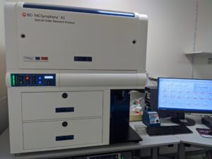
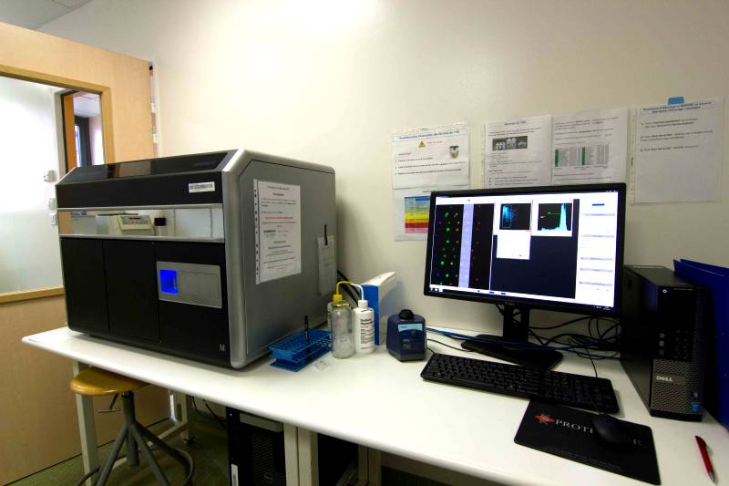 ImageStreamX MarkII