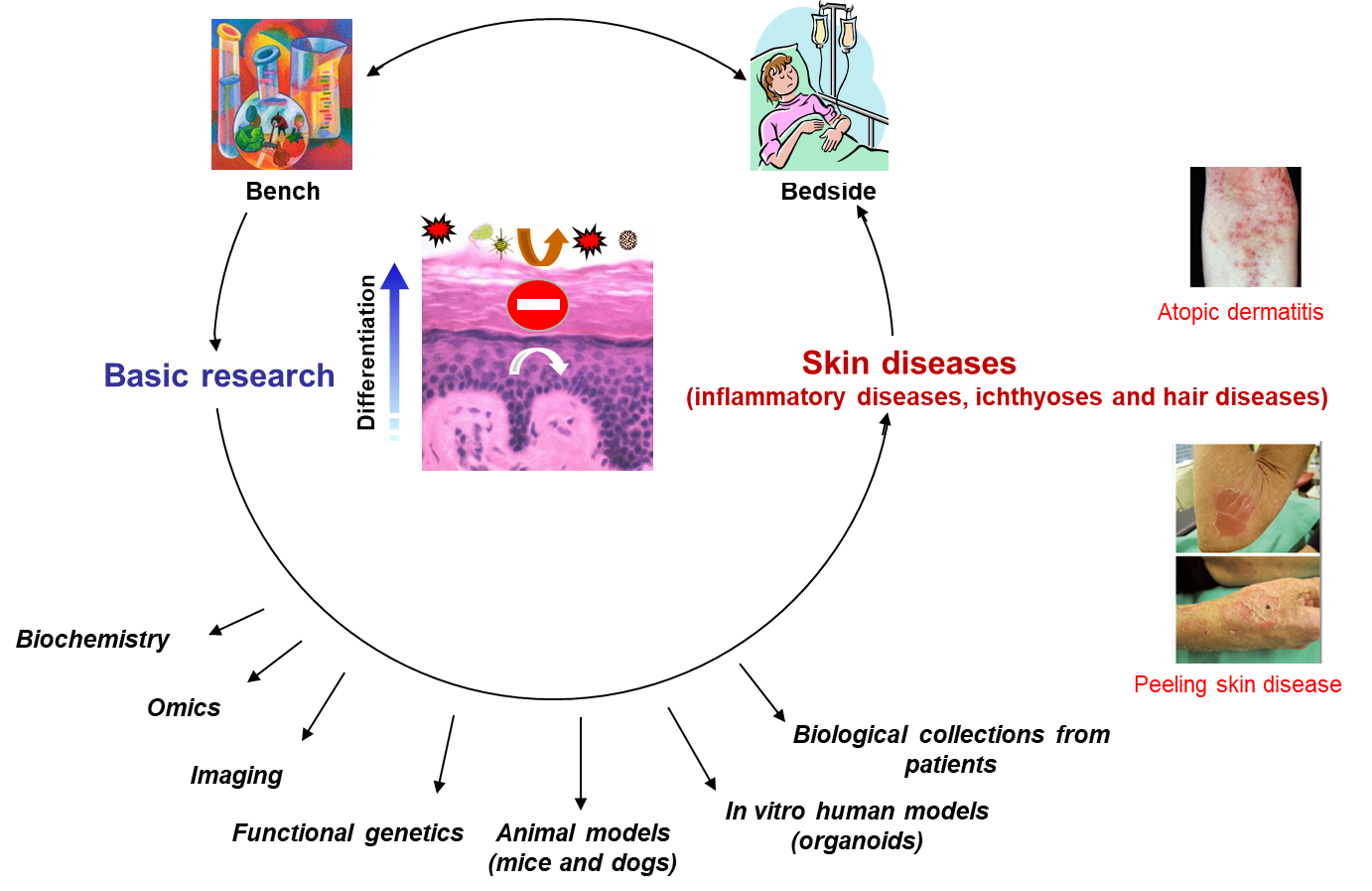
Epidermal barrier and keratinocyte differentiation: from normal skin to skin inflammatory diseases
Coordinator : M. SIMON
Scientific objectives
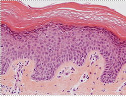
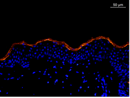
Terminal differentiation of the epidermis is an oriented process during which keratinocytes sequentially turn on and off a series of specific genes in the course of their migration to the external surface of the skin, through the spinous and granular layers. The ultimate step, or cornification, is a genuine process of programmed cell death which results in dramatic structural changes and degradation of the nucleus and cellular organelles, leading to the formation of corneocytes. These cells form the outermost cornified layer of the epidermis. Both the granular and cornified layers allow the epidermis to perform its vital function of multiple barrier between the individual and his environment through their involvement in innate immunity, their high mechanical strength, and their ability to detoxify reactive oxygen species, to limit water loss, to reduce the penetration of UV radiation and to prevent the infiltration of allergens and microorganisms.
From several decades, the objective of our team is to decipher, at the molecular level, the keratinocyte terminal differentiation program, and to know how it is impacted by the environment, and how its impairment induces skin and hair conditions. Thus our work is a continuum between basic research and translational studies. We particularly focus on ichthyoses and inflammatory diseases, including psoriasis and atopic dermatitis. In industrialized countries, the latter affect up to 20% of children and 10% of adults. They therefore represent a huge burden on health care.
Overview of our research
Our Projects
Molecular mechanisms of lamellar body biogenesis and secretion.
The epidermal barrier functions rely on the secretion by granular keratinocytes of the contents of tubulo-vesicular cytoplasmic structures related to lysosomes, derived from the Golgi apparatus, and called lamellar bodies. Lamellar bodies contain various enzymes, including lipases and proteases, anti-microbial peptides, corneodesmosin and lipids. Our aim is to decipher the molecular mechanisms involved in lamellar body biogenesis, trafficking and delivery.
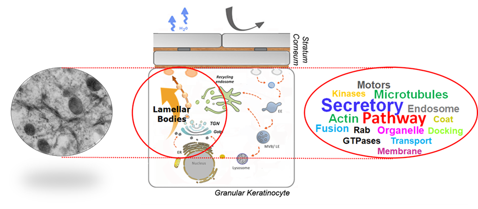
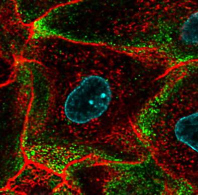
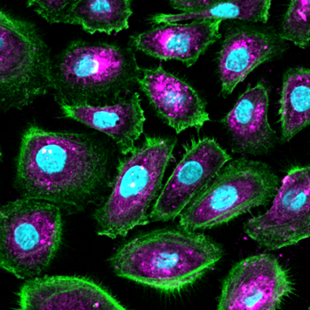
In particular, we investigate the role of Rab family GTPases, cytoskeleton components, as well as the related signaling pathways. In previous reports, we have shown that the Rab11A GTPase and the Myosin5B actin-dependent motor are crucial for lamellar body biogenesis and trafficking. In our ongoing work, we take profit of a unique in vitro experimental model, 3D reconstructed human epidermis where gene modulation can be performed: either down regulation by using RNA interference or overexpression of mutant sequences by using vectors allowing granular keratinocyte-specific expression. Our biological read-out is currently based on biochemical studies, photonic and electronic imaging, and functional assays.
Coordinator: Dr Corinne Leprince
Lipid metabolism and epidermal barrier: from the identification of mutated genes in Autosomal Recessive Congenital Ichthyoses to the patients’ treatment.
Inherited ichthyoses are rare monogenic diseases (prevalence 13.3 per million people in Europe) that occur at birth. They cause abnormal thickening of the skin, dryness, scaling, redness, itching and painful skin fissures involving life-long disfigurement and social ostracism. All forms of ichthyosis lead to a defective epidermal barrier. Recent researches on genes causing Autosomal Recessive Congenital Ichthyoses (ARCI) highlighted the essential role of lipids from the stratum corneum, in particular ceramides, in the epidermal permeability barrier.
Our aim is to better understand the molecular basis of this lipid-based permeability barrier in healthy skin and its impairment in ichthyoses. We are studying enzymes involved in the metabolism of skin ceramides and hope to decipher their subcellular localization and regulation. We particularly focus on PNPLA1 and LIPN, encoded by two ARCI-causing genes.
Another part of our goal is the pre-clinical development of targeted therapies to treat ichthyoses. The topical replacement of the missing lipid is a promising approach to improve the defective permeability barrier encountered in ARCI. We are developing, in an international multidisciplinary collaboration with chemists and experts in skin lipids, innovative substitution systems based on skin-identical lipids encapsulated in nanostructured formulations that will be validated for rescuing the epidermal barrier using pre-clinical in vitro and animal models of ichthyosis.
We also closely collaborate with dermatologists from the South Reference Center for Rare Skin and Mucous Membrane Diseases (recruitment of patients, clinical expertise in ichthyosis) and biologists (molecular diagnosis) at the Toulouse University Hospital. We have access to a large collection of biological samples from patients with ichthyosis and perform clinical and genetic studies to expand the knowledge of the molecular bases of ARCI and help to better define the patient’s characteristics. We also aim to identify undescribed ARCI-causing genes and potentially novel actors involved in lipid metabolism by combine approaches using next generation sequencing and bio clinical analyses.
To achieve all these projects, we benefit from our longstanding experience in the analysis of the epidermal barrier at the functional and molecular levels and we are currently developing high-performance organotypic models of ichthyoses by genome editing and 3D-culture of human keratinocytes.
Coordinator: Dr Nathalie Jonca
Atopic Dermatitis: pathophysiology, epidermal barrier dysfunctions and treatments.
Atopic Dermatitis (AD) pathogenesis results from complex interactions between genetic, immunological and environmental factors. But the primary defect that drives this type 2 immune disease remains controversial. A major discovery has been that loss-of-function mutations in FLG gene are the strongest known genetic risk factor for AD. Indeed, FLG encodes filaggrin, an epidermal protein specifically expressed by differentiated keratinocytes, and essential for the epidermal barrier function. This strongly indicates that skin barrier defects play a key role in the disease, enhancing the penetration of allergens and microbes through the epidermis and systemic IgE sensitization. Our aim is to better understand the relationship between the epidermal barrierdysfunction and the environmental factors in AD, and how this may help to identify new treatments. More specifically:
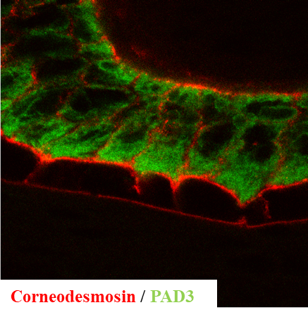
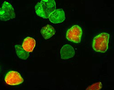
1) Using biochemical in vitro assays, reconstructed human epidermis, and mouse models, we focus on the enzymes (proteases, peptidyl-arginine deiminases (PADs), etc.) involved in the complex metabolism of filaggrin and of other proteins of the S100-fused type protein family, some of them being also associated with epidermal barrier dysfunction in AD. We study how keratinocytes adapt their filaggrin metabolism to the external environment, in particular the relative humidity level.
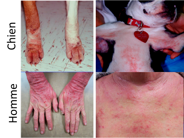 2) Pr Marie-Christine Cadiergues from the Dermatology Department of the National Veterinary School of Toulouse and Michel Simon detail the canine AD at the molecular level, since they believe that the dog disease may be a good model for the human one. Indeed, human and canine AD share many clinical characteristics, in particular spontaneous occurrence. They also develop canine skin models, to confirm pathophysiological hypotheses and test therapeutics, both for veterinary and human medicine.
3) In collaboration with the Ophthalmology and Dermatology Departments of Toulouse Hospital and the National Reference Center for Keratoconus, we have the opportunity i) to investigate, combining transcriptomic analysis and clinical characterization, why the development of severe conjunctivitis is the major adverse effect of Dupilumab, an anti-IL4/13 receptors used to treat AD patients; and ii) to test the hypothesis that corneal epithelium defects are involved in Keratoconus pathogenesis (a rare disease whose frequency is, for unknown reason, strongly enhanced in AD patients) and not only a consequence of stromal deformation.
4) In a multi-disciplinary (chemists, cell biologists and immunobiologists) collaborative work with R Poupot (Infinity Institute) and two other CNRS teams in Toulouse, we test the possibility to use original anti-inflammatory dendrimers as drugs for topical treatment of inflammatory skin diseases including severe forms of AD and psoriasis.
Coordinator: Dr Michel Simon
Other information
Members
Publications
2025 |
Akiyama, Masashi; Choate, Keith; Hernández-Martín, Ángela; Aldwin-Easton, Mandy; Bodemer, Christine; Gostyński, Antoni; Hovnanian, Alain; Ishida-Yamamoto, Akemi; Malovitski, Kiril; O'Toole, Edel A; Paller, Amy S; Schmuth, Matthias; Schwartz, Janice; Sprecher, Eli; Teng, Joyce M C; Tournier, Céline Granier; Mazereeuw-Hautier, Juliette; Tadini, Gianluca; Fischer, Judith Nonsyndromic epidermal differentiation disorders: a new classification toward pathogenesis-based therapy Journal Article In: Br J Dermatol, vol. 193, no. 4, pp. 619–641, 2025, ISSN: 1365-2133. @article{pmid40308026,Epidermal differentiation disorders (EDDs) encompass inherited conditions characterized by abnormal epidermal differentiation, including nonsyndromic and syndromic subtypes with more extensive cutaneous involvement or palmoplantar keratoderma. Nonsyndromic EDDs (nEDDs) are defined as disorders that primarily affect large areas of skin and adnexal structures without alterations in extracutaneous tissues resulting from the underlying genetic change. To facilitate the development of targeted therapies and to provide clinicians with clearer therapeutic guidance, we have developed a new nomenclature for EDDs that includes the causative altered gene and the nEDD subgroup designation, sometimes with a clinical or histological descriptor or acronym. Historically, many nEDDs have been named on the basis of phenotypic characteristics or associations that are now considered outdated or inappropriate. For example, the term 'harlequin ichthyosis' evokes potentially stigmatizing images. Similarly, the word 'ichthyosis' is derived from the Greek ichthys, meaning fish, and the Greek hystrix, meaning porcupine, further emphasizing the need to abandon derogatory terminology. As a result, the clinical relevance of the previous classification, which included eponymous and/or descriptive titles, has diminished. In the new, gene-based classification, old terms considered pejorative, such as ichthyosis, vulgaris, hystrix and harlequin have been eliminated and eponyms have been replaced. Among the 53 genetically distinct nEDDs are conditions formerly known as autosomal recessive congenital ichthyosis, erythrokeratodermia variabilis et progressiva, Hailey-Hailey disease and Darier-White disease. This review outlines the updated nomenclature and classifications of nEDDs, linked to detailed clinical descriptions and representative photographs to guide practitioners. |
Hernández-Martín, Ángela; Paller, Amy S; Sprecher, Eli; Akiyama, Masashi; Mazereeuw-Hautier, Juliette Proposing an immune-inclusive lens to the new epidermal differentiation disorders classification: reply from authors Journal Article In: Br J Dermatol, vol. 193, no. 4, pp. 800–802, 2025, ISSN: 1365-2133. @article{pmid40560203, |
Paller, Amy S; Akiyama, Masashi; Hernández-Martín, Ángela; Mazereeuw-Hautier, Juliette; Sprecher, Eli New gene-based classification of ichthyoses and palmoplantar keratodermas: Hereditary epidermal differentiation disorders Miscellaneous 2025, ISSN: 1097-6787. @misc{pmid40907761, |
Zingkou, Eleni; Reynier, Marie; Pampalakis, Georgios; Serre, Guy; Jonca, Nathalie; Sotiropoulou, Georgia Deletion of the Epidermal Protease Aggravates the Symptoms of Congenital Ichthyosis -nEDD Journal Article In: Int J Mol Sci, vol. 26, no. 17, 2025, ISSN: 1422-0067. @article{pmid40943523,Congenital ichthyoses, now grouped under the acronym EDD (Epidermal Differentiation Disorders), include nonsyndromic forms (nEDD) that may be caused by loss-of-function mutations in the gene encoding corneodesmosin (-nEDD, formerly Peeling skin syndrome type 1). It is characterized by skin peeling, inflammation, itching and food allergies, while no specific therapy is currently available. High levels of KLK5, the serine protease that initiates the desquamation cascade, are found in the epidermis of -nEDD patients. Thus, we hypothesized that KLK5 inhibition would alleviate the symptoms of -nEDD and could serve as a new pharmacological target. A human epidermal equivalent (HEE) model for -nEDD was developed using shRNA-mediated knockdown. This model was characterized and used to assess the role of KLK5 knockdown on -nEDD. Also, mice were crossed with mice, the murine model of -nEDD, to examine in vivo the effect(s) of deletion in -nEDD. Both models recapitulated the -nEDD desquamating phenotype. Elimination of KLK5 aggravated the -nEDD phenotype. Epidermal proteolysis was surprisingly elevated, while severe ultrastructural (corneo)desmosomal alterations increased epidermal barrier permeability and detachment was manifested. Based on these results, we concluded that targeting epidermal proteolysis with ablation cannot compensate for the loss of corneodesmosin and rescue over-desquamation of the -nEDD. Possibly, in the absence of KLK5, other proteases take over which increases the severity of over-desquamation in . The translational outcome is that over-desquamation may not always be rescued by eliminating epidermal proteolysis, but fine protease modulation is more likely required. |
Hernández-Martín, Ángela; Paller, Amy S; Sprecher, Eli; Akiyama, Masashi; Tournier, Céline Granier; Aldwin-Easton, Mandy; Bodemer, Christine; Choate, Keith; Fischer, Judith; Gostynski, Antoni; Hovnanian, Alain; Ishida-Yamamoto, Akemi; O'Toole, Edel A; Schmuth, Matthias; Schwartz, Janice; Tadini, Gianluca; Teng, Joyce; Mazereeuw-Hautier, Juliette A proposal for a new pathogenesis-guided classification for inherited epidermal differentiation disorders Journal Article In: Br J Dermatol, vol. 193, no. 3, pp. 544–548, 2025, ISSN: 1365-2133. @article{pmid40155206, |
Mazereeuw-Hautier, Juliette; Paller, Amy S; Dreyfus, Isabelle; Sprecher, Eli; O'Toole, Edel; Bodemer, Christine; Akiyama, Masashi; Diociaiuti, Andrea; Hachem, Maya El; Fischer, Judith; Gonzalez-Sarmiento, Rogelio; Gutiérrez-Cerrajero, Carlos; Ott, Hagen; Has, Cristina; Jonca, Nathalie; Tournier, Céline Granier; Milesi, Sarah; Texier, Hélène; Martinez, Ana; Traupe, Heiko; Salavastru, Carmen Maria; Schmuth, Matthias; Giehl, Kathrin; Aldwin, Mandy; Morales, Ruth Anton; Santos, Saturnino; Morren, Marie-Anne; Audouze, Anne; Malhotra, Raman; Veldman, Karin; Narbutt, Joanna; Süßmuth, Kira; Hernandez-Martin, Angela; Gostynski, Antoni Management of congenital ichthyoses: guidelines of care: Part one: 2024 update Journal Article In: Br J Dermatol, vol. 193, no. 1, pp. 16–27, 2025, ISSN: 1365-2133. @article{pmid40156154,In 2019, a group of experts published the first European guidelines for the management of congenital ichthyoses after a multidisciplinary expert meeting held in 2016. An update of these guidelines and literature search was planned every 5 years, given the clinical, molecular and therapeutic advances, including the use of biologic therapies. We present here updated guidelines that have been developed by a reorganized multidisciplinary group of international experts. The evidence is based on a systematic review of recent literature, discussions and consensus reached at an expert conference held in June 2023. The guidelines provide summarized evidence and expert-based recommendations that aim to guide clinicians in the management of these rare and often complex diseases. These guidelines consist of two sections. This Part one covers topical and systemic therapies (including oral retinoids, biologics and Janus kinase inhibitors), future therapeutic approaches, psychosocial management, telemedicine, communicating the diagnosis and genetic counselling, prenatal diagnosis and preimplantation genetic testing. |
Mazereeuw-Hautier, Juliette; Paller, Amy S; O'Toole, Edel; Dreyfus, Isabelle; Bodemer, Christine; Akiyama, Masashi; Diociaiuti, Andrea; Hachem, Maya El; Fischer, Judith; Gonzalez-Sarmiento, Rogelio; Gutiérrez-Cerrajero, Carlos; Ott, Hagen; Has, Cristina; Jonca, Nathalie; Tournier, Céline Granier; Martinez, Ana; Traupe, Heiko; Salavastru, Carmen Maria; Schmuth, Matthias; Sprecher, Eli; Giehl, Kathrin; Aldwin, Mandy; Morales, Ruth Anton; Santos, Saturnino; Morren, Marie-Anne; Audouze, Anne; Malhotra, Raman; Veldman, Karin; Narbutt, Joanna; Süßmuth, Kira; Gostynski, Antoni; Hernandez-Martin, Angela Management of congenital ichthyoses: guidelines of care: Part two: 2024 update Journal Article In: Br J Dermatol, vol. 193, no. 1, pp. 28–43, 2025, ISSN: 1365-2133. @article{pmid40190069,In 2019, a group of experts published the first European guidelines for the management of congenital ichthyoses after a multidisciplinary expert meeting held in 2016. An update of these guidelines and literature search was planned every 5 years, given the clinical, molecular and therapeutic advances, including the use of biologic therapies. We present here updated guidelines that have been developed by a reorganized multidisciplinary group of international experts after a systematic review of recent literature, discussions and consensus reached at an expert conference held in June 2023. The guidelines provide summarized evidence and expert-based recommendations that aim to guide clinicians in the management of these rare and often complex diseases. These guidelines consist of two sections. Part one is reported elsewhere. Here, Part two covers the management of complications (eye, ear-nose-throat, pruritus, pain, cutaneous infections, vaccinations, growth failure and nutritional deficiency, hair and nail anomalies, reaction to hot and cold climates, physical limitations, comorbidities) and the particularities of the neonatal period and Netherton syndrome. |
Pons, Carole; Lachambre, Simon; Goudouneche, Dominique; Simon, Michel; Leprince, Corinne Rab27B GTPase Regulates Late Steps of Lamellar Body Trafficking Journal Article In: J Invest Dermatol, 2025, ISSN: 1523-1747. @article{pmid40473201, |
Mazereeuw-Hautier, J Epidermolytic ichthyosis: New insights and ongoing challenges Journal Article In: J Eur Acad Dermatol Venereol, vol. 39, no. 5, pp. 893–894, 2025, ISSN: 1468-3083. @article{pmid40277215, |
Roux, Solène; Marchès, Aurélie; Galiacy, Stéphane; Merbahi, Nofel; Simon, Michel Biological solutions activated by cold plasma at atmospheric pressure: A new therapeutic approach for skin wound healing Journal Article In: Biomed Pharmacother, vol. 186, pp. 118001, 2025, ISSN: 1950-6007. @article{pmid40138920,Chronic wounds are a major public health problem, and nearly 35 % of them do not heal with conventional treatments. The direct application of cold plasmas at atmospheric pressure, partially ionized gases, is an emerging technology with a range of potential biomedical applications, including the improvement of wound healing. A new method that is easier to implement has been developed: the use of biological solutions exposed to cold plasmas at atmospheric pressure, known as plasma-activated media (PAM). Numerous preclinical studies and in vitro models indicate that PAM treatments facilitate wound healing by promoting the migration of cell types such as keratinocytes, fibroblasts and mesenchymal stem/stromal cells, stimulating angiogenesis, and inhibiting bacterial proliferation, all of which are critical to this vital process. PAM treatments modulate the inflammatory response, induce the expression of growth factors and matrix metalloproteinases, reduce cellular adhesion, promote cytoskeletal modifications and activate several biochemical pathways involved in the wound healing process, possibly through the action of plasma-generated reactive oxygen and nitrogen species. Some studies have shown that PAM may have applications in the treatment of other skin conditions either by reducing the production of pro-inflammatory cytokines or by inducing apoptosis of tumor cells. PAM treatments therefore represent a promising new therapy for the management of dermatological conditions, particularly for chronic skin and mucosal wounds. |
Cassagne, Myriam; Galiacy, Stéphane; Kychygina, Anna; Chapotot, Eric; Wallaert, Martin; Vabres, Bertrand; Tauber, Marie; Barbarot, Sébastien; Paul, Carle; Fournié, Pierre; Simon, Michel In: J Invest Dermatol, vol. 145, no. 5, pp. 1050–1059.e6, 2025, ISSN: 1523-1747. @article{pmid39306032,Dupilumab has demonstrated efficacy in the treatment of atopic dermatitis. However, a subset of patients experiences ocular adverse events (OAEs), including conjunctivitis and dry eye syndrome, the pathological mechanisms of which are still unknown. In a bicentric study, we used DNA microarray analysis to compare the transcriptome of conjunctival cells of patients with atopic dermatitis collected by impression cytology before (M0) and 4 months after (M4) initiating dupilumab treatment. Thirty-six patients were included and divided in 2 groups according to their ophthalmological status at M4: 12 with OAEs (OAE+) and 24 without (OAE-). The analysis revealed 52 differentially expressed genes between OAE+ and OAE- patients at M0 and 113 at M4. Ingenuity Pathway Analysis enrichment revealed a psoriasis signature in OAE+ patients, both before and after OAE outcomes. In addition, we noticed the overexpression of several genes involved in keratinocyte differentiation, particularly encoding cornified envelope components. Among the 16 differentially expressed genes selected for real-time RT-PCR validation, 9 were confirmed as upregulated at M4 in OAE+ versus OAE- patients, validating the psoriasis signature, whereas MUC7 was downregulated. In conclusion, these results suggest that a conjunctival transcriptomic profile predisposes some patients with atopic dermatitis to developing OAEs upon dupilumab treatment. |
Combarros, Daniel; Brahmi, Rahma; Musaefendic, Emma; Heit, Alizée; Kondratjeva, Jevgenija; Moog, Fabien; Pressanti, Charline; Lecru, Line A; Arbouille, Sabine; Laffort, Catherine; Goudounèche, Dominique; Brun, Jessie; Simon, Michel; Cadiergues, Marie-Christine In: JID Innov, vol. 5, no. 2, pp. 100330, 2025, ISSN: 2667-0267. @article{pmid39811760,Our objectives were to explore epidermal barrier defects in dogs with atopic dermatitis and to determine whether the defects are genetically determined or secondary to skin inflammation. First, the expression of filaggrin, corneodesmosin, and claudin1, analyzed using indirect immunofluorescence in skin biopsies collected from 32 healthy and 32 dogs with atopic dermatitis, was weaker in the atopic skin ( .003). Second, primary keratinocytes of atopic dogs and healthy dogs were used to produce 3-dimensional reconstructed canine epidermis. The expression of the same proteins was analyzed using indirect immunofluorescence, immunoblotting, and RT-qPCR, whereas reconstructed canine epidermis morphology was investigated by transmission electron microscopy, and the barrier was investigated by functional assays. Next, inflammatory cytokines (IL-4, IL-13, IL-31, and TNFα) were added to the culture medium. The morphology, protein expression, and barrier function of the reconstructed canine epidermis were similar whether produced with keratinocytes from healthy dogs or dogs with atopy. Addition of inflammatory cytokines impaired the protein expression and epidermal barrier of the 2 types of reconstructed canine epidermis equally. To conclude, the reduced expression of epidermal barrier proteins observed in vivo was not reproduced in vitro unless cytokines were used, suggesting that it is induced by the inflammatory milieu. |
Morice, Camille; Saunier, Valentine; Kyheng, Maeva; Fournie, Pierre; Touboul, David Meibomian gland atrophy in Keratoconus: A case-control study Journal Article In: Eur J Ophthalmol, pp. 11206721251327651, 2025, ISSN: 1724-6016. @article{pmid40123225,PurposeTo assess the Meibomian Gland Atrophy (MGA) in keratoconus patients by comparison to healthy controls.SettingFrench Keratoconus national reference center, Bordeaux, France.DesignObservational, monocentric, comparative case control study.MethodsEighty-eight keratoconic eyes free of any surgical treatment (KC group) and 88 healthy eyes (NKC group) matched for age and gender, included from December 2018 to May 2019, underwent infrared meibography providing a meiboscore of MGA (primary outcome measure), scaled from 0 to 6. Secondary outcomes including Lipid Layer Thickness (LLT) assessment using interferometry, Tear Break-Up Time (T-BUT) evaluated by a slit lamp examination, corneal OCT-based topography, and two dry eye questionnaires (OSDI and SPEED) filled by the patients during the consultation.ResultsA significant difference in distribution of meiboscore scores between groups ( < 0.0001 for total, superior and inferior meiboscore) was found, with lower scores in controls compared to eyes with keratoconus. Using a cut-off value of ≥2 as abnormal meiboscore, 64.8% of eyes of cases had abnormal meiboscore by comparison to only 27.3% of controls (OR, 4.91; 95%CI, 2.56 to 9.38). Patients with keratoconus had higher OSDI (mean difference 15.3, 95%CI 1.8 to 1.7) and SPEED (mean difference 2.5, 95%CI 2.5 to 4.5) results. No significant difference was found in T-BUT whereas keratitis was more common in cases.ConclusionKeratoconus was associated with higher meibomian gland atrophy in this study. |
Paul, Carle; Stratigos, Alexander J; Nijsten, Tamar; Gisondi, Paolo; Salavastru, Carmen; Röecken, Martin; Taieb, Charles; Sampogna, Francesca; Trakatelli, Myrto; Puig, Luis; Richard, Marie A The Journal Article In: Minerva Med, vol. 116, no. 1, pp. 81–83, 2025, ISSN: 1827-1669. @article{pmid38743041, |
Dumitrache, Mirabela Oana; Ursache, Aurora Livia; Toma, Corina; Negoescu, Andrada; Rietmann, Stefan Jonas; Leeb, Tosso; Cadiergues, Marie-Christine Canine exfoliative cutaneous lupus erythematosus in two mixed breed littermates Journal Article In: Vet Dermatol, vol. 36, no. 1, pp. 99–103, 2025, ISSN: 1365-3164. @article{pmid39344864,Canine exfoliative cutaneous lupus erythematosus (ECLE) is the rarest variant of cutaneous lupus in dogs and has strong breed predilections. This report presents the clinical, histopathological and immunohistochemical features of two ECLE cases in mixed breed littermates and confirms the expected genetic mutation. A therapeutic response to oclacitinib also is documented. |
Bernard, Pauline; Pell, Nuria; Mazereeuw-Hautier, Juliette; Jonca, Nathalie Novel ABCA12 Missense Variant in a Patient with Congenital Ichthyosis and Palmoplantar Keratoderma Journal Article In: Acta Derm Venereol, vol. 105, pp. adv42502, 2025, ISSN: 1651-2057. @article{pmid39749396, |
Leprince, Corinne; Simon, Michel Epidermal lamellar bodies, essential organelles for the skin barrier Journal Article In: Front Cell Dev Biol, vol. 13, pp. 1597884, 2025, ISSN: 2296-634X. @article{pmid40698039b,Skin lamellar bodies are members of the Lysosome-Related-Organelle (LRO) family, characterized by specific features related to the skin's primary function, i.e., protecting the body from external assaults while minimizing dehydration. In the uppermost living cell layers of the epidermis, the vesicles and tubulovesicular network that make up the « lamellar body system » as identified by electron microscopists, play a crucial role in maintaining the skin barrier. As a secretory compartment, lamellar bodies carry a variety of compounds that, when released in the extracellular space or exposed at the membrane, contribute to the unique hydrophobic structure of the upper epidermis (lipids and lipid metabolism enzymes), regulate desquamation (proteases and inhibitors) and provide anti-microbial defense. The molecular machinery involved in the biogenesis and trafficking of skin lamellar bodies is only beginning to be deciphered, including the Rab11A GTPase, the Myosin5B molecular motor, and the CHEVI complex. This later one is constituted of the Vps33B and VIPAR tethering molecules, whose mutations lead to the ARC and ARKID syndromes. Further studies are needed to identify the key molecules regulating the various stages of LB biogenesis, maturation and exocytosis. It is likely that some of these molecules will be shared with other members of the LRO family. These studies will further enhance our understanding of the relationships between lamellar body trafficking and skin barrier dysfunction. |
Briot, Julie; Pons, Carole; Foucher, Aude; Goudounèche, Dominique; Gaudenzio, Nicolas; Donovan, Mark; Bernard, Dominique; Méchin, Marie-Claire; Simon, Michel Prolyl Endopeptidase Is Involved in Filaggrinolysis and Cornification Journal Article In: J Invest Dermatol, vol. 145, no. 1, pp. 98–108.e15, 2025, ISSN: 1523-1747. @article{pmid38879153,FLG is a well-known biomarker of atopic dermatitis and skin dryness. Its full proteolysis (or filaggrinolysis) produces the major constituents of the natural moisturizing factor. Some proteases/peptidases remain to be identified in this multistep process. Mining 16 omics analyses, we identified prolyl endopeptidase (PREP) as a candidate peptidase. Indirect immunofluorescence and confocal analysis demonstrated its localization in the granular and deep cornified layers, where it colocalized with FLG. Tandem mass spectroscopy and fluorescent quenching activity assays showed that PREP cleaved several synthetic peptides derived from the FLG sequence, at the carboxyl side of an internal proline. Deimination of these peptides increased PREP enzymatic efficiency. Specific inhibition of PREP in reconstructed human epidermis using benzyloxycarbonyl-pro-prolinal induced the accumulation of FLG monomers. Downregulation of PREP expression in reconstructed human epidermis using RNA interference confirmed the impact of PREP on FLG metabolism and highlighted a more general role of PREP in keratinocyte differentiation. Indeed, quantitative global proteomic, western blotting, and RT-qPCR analyses showed a strong reduction in the expression of bleomycin hydrolase, known to be involved in filaggrinolysis, and of several other actors of cornification such as loricrin. Consequently, at the functional level, the transepidermal electric resistance was drastically reduced. |
2024 |
Pancarte, Mikaël; Leignadier, Julie; Courrech, Séverine; Serre, Guy; Attia, Joan; Jonca, Nathalie Strengthening the Skin Barrier by Using a Late Cornified Envelope 6A-Derived Biomimetic Peptide Journal Article In: Exp Dermatol, vol. 33, no. 10, pp. e15191, 2024, ISSN: 1600-0625. @article{pmid39397370,Changes in the expression of cornified envelope (CE) components are a hallmark of numerous pathological skin conditions and aging, underlying the importance of this stratum corneum structure in the homeostasis of the epidermal barrier. We performed a detailed characterisation of LCE6A, a member of the Late Cornified Envelope protein family. Immunohistochemical and immunoblot experiments confirmed that LCE6A is expressed late during epidermal differentiation. Crosslinking assays of recombinant LCE6A performed either in situ on human skin sections or in vitro demonstrated that LCE6A is indeed a substrate of transglutaminases and crosslinked to CEs. LCE6A-derived peptides containing a glutamine-lysine sequence retained these properties of the full-length protein and reinforced the mechanical resistance of CE submitted to sonication. We designed P26, a LCE6A-derived biomimetic peptide that similarly reinforced CE in vitro, and evaluated its protective properties ex vivo, on human skin explants, and in two double blind and vehicle-controlled clinical trials. P26 was able to protect the skin from barrier disruption, to limit the damage resulting from a defective barrier, and could improve the signs of aging such as loss of skin firmness and increased skin roughness. Hence, our detailed characterisation of LCE6A as a component of the CE enabled us to develop a LCE6A-derived peptide, biologically active with a new and original mode of action that could be of great interest as a cosmetic ingredient and a pharmacologic agent. |
Mallet, Stéphanie; Frankel, Diane; Jonca, Nathalie; Cano, Aline; Roll, Patrice; Kaspi, Elise Leukocytes containing lipid inclusions in congenital ichthyosis without classical Chanarin-Dorfman mutations Miscellaneous 2024, ISSN: 1365-4632. @misc{pmid38581117, |
Dumitrache, M O; Kalmár, Z; Roumegous, S; Charmes, K; Delverdier, M; Net, J L Le; Cadiergues, M C Feline straelensiosis: Clinical and histopathological description of a case and first genetic characterisation of Straelensia cynotis Journal Article In: Vet Dermatol, vol. 35, no. 2, pp. 234–237, 2024, ISSN: 1365-3164. @article{pmid37953433,Straelensia cynotis is a trombidioid mite that causes painful, usually nonpruritic nodular dermatitis mainly in the dorsal region of dogs. This case report describes the first observation of feline straelensiosis in Europe with clinicopathological findings. Molecular characterisation of the parasite was performed and compared with mites collected from dogs. |
Simon, Michel Newly Discovered Corneodesmosin Defects in Generalized Pustular Psoriasis Journal Article In: J Invest Dermatol, vol. 144, no. 4, pp. 731–733, 2024, ISSN: 1523-1747. @article{pmid38300198, |
Severino-Freire, M; Tournier, C Granier; Chiaverini, C; Audouze, A; Morice-Picard, F; Texier, H; Dreyfus, I; Bing-Lecointe, A-C; Mallet, S; Bodemer, C; Fischer, J; Jonca, N; Mazereeuw-Hautier, J French national protocol for the management of congenital ichthyosis Journal Article In: Ann Dermatol Venereol, vol. 151, no. 1, pp. 103247, 2024, ISSN: 0151-9638. @article{pmid38513308,Congenital ichthyoses (CI) comprise a heterogeneous group of monogenic genetic skin diseases characterized by diffuse scaling, often associated with skin inflammation. Diagnosis of the individual form of ichthyosis is complex and is guided by clinical expertise. CI usually has a major impact on quality of life (QOL) and thus requires lifelong treatment. To date, there are no curative therapies, although various symptomatic treatment options exist. The present protocol for the management of CI has been drawn up in accordance with the recommendations published in 2012 by the French National Authority for Health, based on a literature review, with the help and validation of members of the French network for rare skin diseases (FIMARAD). It provides a summary of evidence and expert-based recommendations and is intended to help clinicians with the management of these rare and often complex diseases. |
Briot, Julie; Arbey, Eric; Goudounèche, Dominique; Bernard, Dominique; Simon, Michel; Méchin, Marie-Claire Human filaggrin monomer does not seem to be a proteasome target Journal Article In: Exp Dermatol, vol. 33, no. 1, pp. e14772, 2024, ISSN: 1600-0625. @article{pmid36807394,Absence of a functional proteasome in the suprabasal layers of the epidermis is responsible for keratosis linearis with ichthyosis congenital and sclerosing keratoderma syndrome. Patient epidermis shows hypergranulosis associated with abnormally shaped keratohyalin granules and abnormal distribution of filaggrin in the Stratum granulosum and Stratum corneum. This suggests that the proteasome is involved in the degradation of filaggrin. To test this hypothesis, the proteasome proteolytic activity was inhibited in 3D reconstructed human epidermis (RHE) with the specific clasto-lactacystin β-lactone inhibitor. Confirming the efficacy of inhibition, ubiquitinated proteins accumulated in treated RHEs as compared to controls. Levels of urocanic acid (UCA) and pyrrolidone carboxylic acid (PCA), the end products of filaggrin degradation, were reduced. However, neither filaggrin accumulation nor appearance of filaggrin-derived peptides were observed. On the contrary, the amount of filaggrin was shown to decrease, and a similar tendency was observed for profilaggrin, its precursor. Accumulation of small cytoplasmic vesicles associated with a significant increase in autophagy markers indicated activation of the autophagy process upon proteasome inhibition. Taken together, these results suggest that the perturbation of UCA and PCA production after proteasome inhibition was probably due to down-regulation of filaggrin expression rather than to blocking of filaggrin proteolysis. |
2023 |
Caengprasath, Natarin; Nizon, Mathilde; Panchaprateep, Ratchathorn; Cogne, Benjamin; Cuinat, Silvestre; Auburt, Hélène; Jonca, Nathalie; Porntaveetus, Thantrira; Shotelersuk, Vorasuk Two novel MBTPS2 missense mutations impairing S2P proteolytic activity lead to IFAP syndrome with new phenotypic anomalies Miscellaneous 2023, ISSN: 1873-569X. @misc{pmid37923657, |
Méchin, Marie-Claire; Simon, Michel Deimination in epidermal barrier and hair formation Journal Article In: Philos Trans R Soc Lond B Biol Sci, vol. 378, no. 1890, pp. 20220245, 2023, ISSN: 1471-2970. @article{pmid37778378,Peptidylarginine deiminases (PADs) transform a protein arginine residue into the non-standard amino acid citrulline. This calcium-dependent post-translational modification of proteins is called citrullination or deimination. As described in this special issue, PADs play a role in various physiological processes, and PAD deregulations are involved in many human diseases. Three PADs are expressed in the epidermis, where their roles begin to be deciphered. PAD1 and PAD3 are involved in keratinocyte differentiation, particularly in the epidermal barrier function, keratins, filaggrin and filaggrin-related proteins being the most abundant deiminated epidermal proteins. Reduced amounts of deiminated proteins and PAD1 expression may be involved in the pathogenesis of psoriasis and atopic dermatitis, two very frequent and chronic skin inflammatory diseases. The trichohyalin/PAD3/transglutaminase three pathway is important for hair shaft formation. Mutations of the gene, leading to a decreased activity or abnormal localization of the corresponding isotype, are the cause of a rare hair disorder called uncombable hair syndrome, and are associated with the central centrifugal cicatricial alopecia, a frequent alopecia mainly affecting women of African ancestry. This article is part of the Theo Murphy meeting issue 'The virtues and vices of protein citrullination'. |
Challamel, C; Hernandez-Martin, A; Tchitchiama, C; Jonca, N; Rossel, S V J; Gostyński, A; Mazereeuw-Hautier, J Patients with autosomal recessive congenital ichthyosis present a distinctive pattern of alopecia Miscellaneous 2023, ISSN: 1468-3083. @misc{pmid37306217, |
Valette, C; Jonca, N; Fischer, J; Pernin-Grandjean, J; Tournier, C Granier; Diociaiuti, A; Neri, I; Dreyfus, I; Furman, M; Giehl, K; Wollenberg, A; Mallet, S; Martin, L; Martin-Santiago, A; Onnis, G; Broue, P; Leclerc-Mercier, S; Schmuth, M; Sprecher, E; Gruber, R; Suessmuth, K; Bourrat, E; Komlosi, K; Hill, S; O'Toole, E A; Schischmanoff, O; Caux, F; Mazereeuw-Hautier, J A retrospective study on the liver toxicity of oral retinoids in Chanarin-Dorfman syndrome Miscellaneous 2023, ISSN: 1468-3083. @misc{pmid37257069, |
Dumitrache, Mirabela O; Cadiergues, Marie-Christine The most effective systemic treatment in dogs with sarcoptic mange: a critically appraised topic Journal Article In: BMC Vet Res, vol. 19, no. 1, pp. 189, 2023, ISSN: 1746-6148. @article{pmid37798627,BACKGROUND: Sarcoptic mange is a common, pruritic parasitic skin disease of dogs. Due to its highly contagious character, it represents a potential veterinary and public health risk. Because of clinical similarity with other diseases, cross-antigenicity, and low sensitivity of available diagnostic methods, therapeutical trial is frequently used to confirm the disease. Considering the variety of available acaricidal molecules as well as the need to use the most effective treatment, the present paper reviews evidence comparing different types of systemic treatment of canine scabies.nnRESULTS: Analysis of the results showed that afoxolaner, fluralaner and sarolaner as well as several macrocyclic lactones such as selamectin, moxidectin and milbemycin oxime can lead to parasitological and clinical cure.nnCONCLUSION: The similarity in the clinical and parasitological efficacy of these substances enhances the need for comparative studies, which could allow the identification of the most efficacious product. |
Jonca, Nathalie; Simon, Michel The Cornified Envelope: A Versatile Contributor to the Epidermal Barrier Journal Article In: J Invest Dermatol, vol. 143, no. 8, pp. 1335–1337, 2023, ISSN: 1523-1747. @article{pmid37149811, |
Alioli, Adebayo Candide; Briot, Julie; Pons, Carole; Yang, Hang; Gairin, Marie; Goudounèche, Dominique; Cau, Laura; Simon, Michel; Méchin, Marie-Claire In: Cell Death Discov, vol. 9, no. 1, pp. 198, 2023, ISSN: 2058-7716. @article{pmid37385992,Deimination is a post-translational modification catalyzed by a family of enzymes named peptidylarginine deiminases (PADs). PADs transform arginine residues of protein substrates into citrulline. Deimination has been associated with numerous physiological and pathological processes. In human skin, three PADs are expressed (PAD1-3). While PAD3 is important for hair shape formation, the role of PAD1 is less clear. To decipher the main role(s) of PAD1 in epidermal differentiation, its expression was down-regulated using lentivirus-mediated shRNA interference in primary keratinocytes and in three-dimensional reconstructed human epidermis (RHE). Compared to normal RHEs, down-regulation of PAD1 caused a drastic reduction in deiminated proteins. Whereas proliferation of keratinocytes was not affected, their differentiation was disturbed at molecular, cellular and functional levels. The number of corneocyte layers was significantly reduced, expression of filaggrin and cornified cell envelope components, such as loricrin and transglutaminases, was down-regulated, epidermal permeability increased and trans-epidermal-electric resistance diminished drastically. Keratohyalin granule density decreased and nucleophagy in the granular layer was disturbed. These results demonstrate that PAD1 is the main regulator of protein deimination in RHE. Its deficiency alters epidermal homeostasis, affecting the differentiation of keratinocytes, especially the cornification process, a special kind of programmed cell death. |
Banuls, Damien; Brun, Jessie; Blua, Jean-Louis; Cadiergues, Marie Christine A Dietary Plant Extract Formulation Helps Reduce Flea Populations in Cats: A Double-Blind Randomized Study Journal Article In: Pharmaceuticals (Basel), vol. 16, no. 2, 2023, ISSN: 1424-8247. @article{pmid37259343,There is a growing demand for natural products to be used to control fleas in pets. A prospective, double-blind, randomized, placebo-controlled study evaluated the efficacy of the biological plant-based food supplement Bioticks (thyme, rosemary, lemon balm, fenugreek, wormwood, and lemongrass extracts) as a flea control product in naturally flea-infested cats with an indoor-outdoor lifestyle. Ten cats were used as placebo controls (group A). Ten other cats were fed the same daily diet but supplemented with Bioticks (group B). Fleas were counted by combing at D0 and D0 + 14 days, then one, two, three, four, and five months after the start of this study. No flea treatment was administered, and no environmental changes were made for six months prior to the start and throughout this study. The product was well-tolerated. The mean flea population in group B progressively and steadily decreased to reach 3.3 ± 2.1 at month five. At the same time and under similar maintenance conditions, the average flea population in group A remained stable (14.3 ± 2.5) until the fifth month. The percentages of efficacy (Abbott formula) in group B compared to group A was 27%, 20%, 52%, 66%, and 77%, respectively, at one, two, three, four, and five months after the start of this study. |
Dumitrache, Mirabela Oana; Györke, Adriana; Julien, Florie; Kondratjeva, Jevgenija; Cadiergues, Marie-Christine Case report: Identification of the tropical rat mite () on a domestic donkey in France Journal Article In: Front Vet Sci, vol. 10, pp. 1141290, 2023, ISSN: 2297-1769. @article{pmid37303734,A 25-year-old donkey was referred for a generalized, pruritic and severe exfoliative dermatitis that had been evolving for several years, with deterioration in the last few months. Close examination of the skin surface revealed numerous small, dark, mobile elements identified as confirmed by DNA sequencing. The severity, type and topography of the lesions called for complementary examinations, leading to a second diagnosis of cutaneous epitheliotropic T-cell lymphoma. The lack of clinical improvement after antiparasitic therapy despite parasite clearance, suggests opportunistic behavior of . To the best of our knowledge, this is the first report of the presence of a tropical rat mite on a donkey, thus expanding the known host spectrum of this zoonotic parasite. Further potential questions concern the implication of this new host as a possible source of human contamination. |
Kondratjeva, Jevgenija; Pressanti, Charline; Reynolds, Brice S; Trumel, Catherine; Delverdier, Maxence; Normand, Anne-Cécile; Soetart, Nicolas; Guillot, Jacques; Cadiergues, Marie Christine Multifocal cutaneous phaeohyphomycosis caused by with clinical resolution in an immunocompromised cat Journal Article In: JFMS Open Rep, vol. 9, no. 1, pp. 20551169231164610, 2023, ISSN: 2055-1169. @article{pmid37123554,CASE SUMMARY: A 3-year-old neutered domestic shorthair cat with a long history of idiopathic immune-mediated haemolytic anaemia and thrombocytopenia treated with ciclosporin and prednisolone was referred 2 months after the appearance of nodular dermatitis. A single pigmented nodule was present in the lateral carpal region of the right foreleg. The lesion was 7 mm in diameter, non-exudative and cutaneous to subcutaneous. Fine-needle aspiration of the mass revealed the presence of pigmented fungal elements. Excisional surgery was planned; in the meantime, a plaque-like lesion developed in the interorbital region. Histopathological examination confirmed the presumptive diagnosis of phaeohyphomycosis, and was identified as the aetiological agent. Itraconazole, given orally at a dose of 10 mg/kg for 8 weeks following surgery, enabled clinical resolution despite continued use of immunosuppressants. The follow-up was carried out over 14 weeks.nnRELEVANCE AND NOVEL INFORMATION: This case report provides the first evidence of multifocal cutaneous phaeohyphomycosis caused by with clinical resolution after combined surgical and itraconazole treatment in an immunocompromised cat. |
Kondratjeva, Jevgenija; Brun, Jessie; Amalric, Nicolas; Moog, Fabien; Combarros, Daniel; Pressanti, Charline; Zemirline, Claudine; Maubert, Nadège; Ollivier, Elodie; Gatellet, Marina; Cadiergues, Marie Christine Performance and Tolerance of a Protocol for Idiopathic Chronic Greasy Seborrhea in 18 Dogs Using a Shampoo and Mousse Containing Plant Extracts Journal Article In: Vet Sci, vol. 10, no. 2, 2023, ISSN: 2306-7381. @article{pmid36851399,The study aimed to evaluate the tolerance, performance and effect on hair lipids and skin hydration of a protocol combining applications of one shampoo and subsequent mousses containing plant extracts (Ophytrium and Seboliance) in dogs with an undiagnosed chronic greasy keratinisation disorder. Six dogs were washed with plain water on day (D)0. Twelve dogs were shampooed on D0 and received eight mousse applications at 48-72 h intervals from D2 to D18. Clinical score (CS), Natural Moisturizing Factors (NMF) and hair lipids (HL) were evaluated on D0, D0 + 4 h, D7, D14 and D24. At baseline, no significant differences were observed in CS, NMF and HL between groups. In the control group, CS and HL remained stable throughout the study while a slight decrease in NMF was observed at D0 + 4 h. CS was significantly reduced in the test group between D0 and D7 (-53%) which reached 91% at D24 ( < 0.05), with no side effects. NMF levels decreased in the test group at D0 + 4 h (-73%, < 0.0001) and returned to baseline from D14. In conclusion, one shampoo and subsequent mousse applications rapidly and safely improved coat quality in dogs with an undiagnosed keratinisation disorder without affecting NMF and HL contents over the study period. |
2022 |
Lupasco, Tatiana; He, Zhiguo; Cassagne, Myriam; Sagnial, Tomy; Brion, Lise; Fournié, Pierre; Gain, Philippe; Thuret, Gilles; Allouche, Michèle; Malecaze, François; Simon, Michel; Galiacy, Stéphane Corneal epithelium in keratoconus underexpresses active NRF2 and a subset of oxidative stress-related genes. Journal Article In: PLoS One, 2022. @article{T2022, |
Marches, Aurélie; Clement, Emily; Albérola, Géraldine; Rols, Marie-Pierre; Cousty, Sarah; Simon, Michel; Merbahi, Nofel In: Int J Mol Sci, 2022. @article{Aurélie2022, |
Padhi, Avinash; Srivastava, Ankit; Ramesh, Abarajitha; Ehrström, Marcus; Simon, Michel; Sonkoly, Eniko; Eidsmo, Liv; Bergman, Peter; Lysell, Josefin In: Invest Dermatol, 2022. @article{Padhi2022, |
Rabionet, Mariona; Bernard, Pauline; Pichery, Mélanie; Marsching, Christian; Bayerle, Aline; Dworski, Shaalee; Kamani, Mustafa; Chitraju, Chandramohan; Gluchowski, Nina; Gabriel, Katlyn; Asadi, Abolfazl; Ebel, Philipp; Hoekstra, Menno; Menno, Sabrina; Ntambi, James; Jacobsson, Anders; Willecke, Klaus; Medin, Jeffrey; Jonca, Nathalie; Sandhoff, Roger In: Lipids, 2022. @article{Mariona2022, |
Basmanav, Buket; Cesarato, Nicole; Kumar, Sheetal; Borisov, Oleg; Kokordelis, Pavlos; Ralser, Damian; Wehner, Maria; Axt, Dais; Xiong, Xong; Thiele, Holger; Dolgin, Vadim; Gossmann, Yasmina; Fricker, Nadine; Dewenter, Malin Katharina; Weller, Karsten; Suri, Mohnish; Reichenbach, Herbert; Oji, Vinsenz; Addor, Marie-Claude; Ramirez, Karla; Stewart, Helen; Bartels, Natalie Garcia; Weibel, Lisa; Wagner, Nicola; George, Susannah; Kilic, Arzu; Tantcheva-Poor, Iliana; Stewart, Alison; Dikow, Nicola; Blaumeiser, Bettina; Medvecz, Marta; Blume-Peytavi, Ulrike; Farrant, Paul; Grimalt, Ramon; Bertok, Sara; Bradley, Lisa; Eskin-Schwartz, Marina; Birk, Ohad Samuel; Bygum, Anette; Simon, Michel; Krawitz, Peter; Fischer, Christine; Hamm, Henning; Fritz, Gunter; Betz, Regina Assessment of the Genetic Spectrum of Uncombable Hair Syndrome in a Cohort of 107 Individuals. Journal Article In: JAMA Dermatol, 2022. @article{Basmanav2022, |
Cadau, Sébastien; Gault, Manon; Berthelemy, Nicolas; Hsu, Chiung-Yueh; Danoux, Louis; Pelletier, Nicolas; Goudounèche, Dominique; Pons, Carole; Leprince, Corinne; André-Frei, Valérie; Simon, Michel; Pain, Sabine An Inflamed and Infected Reconstructed Human Epidermis to Study Atopic Dermatitis and Skin Care Ingredients. Journal Article In: Int J Mol Sci. , 2022. @article{Cadau2022, |
Gouarderes, Sara; Marches, Aurélie; Vicendo, Patricia; Fourquaux, Isabelle; Simon, Michel; Merbahi, Nofel; Gibot, Laure Cold helium plasma jet does not stimulate collagen remodeling in a 3D human dermal substitute. Journal Article In: Bio electrochemistry, 2022. @article{Sara2022, |
Henriet, Elodie; Abdallah, Florence; Laurent, Yoan; Guimpied, Cyril; Clement, Emily; Simon, Michel; Pichon, Chantal; Baril, Patrick Targeting TGF-β1/miR-21 pathway in keratinocytes reveals protective effects of silymarin on imiquimod-induced psoriasis mouse model. Journal Article In: J Invest Dermatol Innov, 2022. @article{Elodie2022, |
MandyJoosten,; Clabbers, Julia; Jonca, Nathalie; Mazereeuw-Hautier, Juliette; Gostyński, Antony New developments in the molecular treatment of ichthyosis: review of the literature. Journal Article In: Orphanet J Rare Dis, 2022. @article{Joosten2022, |
Kychygina, Anna; Cassagne, Myriam; Tauber, Marie; Galiacy, Stéphane; Paul, Carle; Fournié, Pierre; Simon, Michel Dupilumab-Associated Adverse Events During Treatment of Allergic Diseases. Journal Article In: Clin Rev Allergy Immunol. , 2022. @article{Anna2022, |
Moosbrugger-Martinz, Verena; Leprince, Corinne; Méchin, Marie-Claire; Simon, Michel; Blunder, Stefan; Gruber, Robert; Dubrac, Sandrine Revisiting the Roles of Filaggrin in Atopic Dermatitis. Journal Article In: Int J Mol Sci. , 2022. @article{Verena2022, |
Lecru, Line-Alice; Combarros, Daniel; Moog, Fabien; Marinovic, Lukrecija; Kondratjeva, Jevgenija; Amalric, Nicolas; Pressanti, Charline; Cadiergues, Marie-Christine In: Animals (Basel). , 2022. @article{Lecru2022, |
Bruet, Vincent; Mosca, Marion; Briand, Amaury; Bourdeau, Patrick; Pin, Didier; Noelle Cochet-Faivre,; Cadiergues, Marie-Christine Clinical Guidelines for the Use of Antipruritic Drugs in the Control of the Most Frequent Pruritic Skin Diseases in Dogs. Journal Article In: Vet Sci. , 2022. @article{Bruet2022, |
Lasek, Audrey; Bellon, Nathalie; Mallet, Stéphanie; Puzenat, Eve; Bursztejn, Anne-Claire; Abasq, Claire; Mazereeuw-Hautier, Juliette; Chiaverini, Christine; Hubiche, Thomas; Raison-Peyron, Nadia; Thanh, Aurélie Du; Barbarot, Sébastien; Aubert, Hélène; Reguiai, Ziad; Droitcourt, Catherine; Fievet, Charlotte; Bellissen, Astrid; Bachelerie, Marie; Nosbaum, Audrey; Leymarie, Anne; Armingaud, Philippe; Regnault, Marie Masson; Mahé, Emmanuel; of the Société Française de Dermatologie Pédiatrique (GR SFDP), Research Group; of eczéma atopique (GREAT), Research Group In: J Eur Acad Dermatol Venereol., 2022. @article{Audrey2022, |
Debourdeau, Eloi; Planells, Gabriel; Chamard, Chloé; Touboul, David; Villain, Marc; Demoly, Pascal; Babeau, Fanyy; Fournie, Pierre; Daien, Vincent New Keratoconus Risk Factors: A Cross-Sectional Case-Control Study. Journal Article In: J Ophthalmol. , 2022. @article{Eloi2022, |
Crépy, Marie-Noelle; Bensefa-Colas, Linda; Aubin, François; Simon, Michel; Soria, Angèle; research group for atopic dermatitis (Groupe de Recherche sur l’Eczéma Atopique) in collaboration with the French Society of Dermatology, French; of Allergology; French Society of Pediatrics, Venereology; French Society; of Occupational Medicine;, French Society Vocational Guidance for Young Patients with Atopic Dermatitis: A Survey of Physicians' Opinions and Practices. Journal Article In: Acta Derm Venereol. , 2022. @article{Marie-Noelle2022, |
2021 |
Gouarderes, Sara; Marches, Aurélie; Vicendo, Patricia; Fourquaux, Isabelle; Simon, Michel; Merbahi, Nofel; Gibot, Laure Cold atmospheric helium plasma does not stimulate dermal collagen remodeling at tissue scale. Journal Article In: Bioelectrochemistry, 2021. @article{S2021b, |
Luger, Thomas; Amagai, Masayuki; Dreno, Brigitte; Dagnelie, Marie-Ange; Liao, Wilson; Kabashima, Kenji; Schikowski, Tamara; Proksch, Ehrhardt; Elias, Peter; Simon, Michel; Simpson, Eric; Grinich, Erin; Schmuth, Matthias Atopic dermatitis: Role of the skin barrier, environment, microbiome, and therapeutic agents. Journal Article In: Journal of Dermatological Science, 2021. @article{T2021, |
Combarros, Daniel; Goudounèche, Dominique; Cadiergues, Marie-Christine; Simon, Michel The upper epidermis of atopic dogs is altered at the functional and structural levels. Journal Article In: Vet Dermatol, 2021. @article{D2021, |
Hotz, Alrun; Kopp, Julia; Bourrat, Emmanuelle; Oji, Vinzenz; Komlosi, Katalin; Giehl, Kathrin; Bouadjar, Bajar; AnetteBygum,; Tantcheva-Poor, Iliana; Pigg, Maritta Hellström; Has, Cristina; Yang, Zhou; Irvine, Alan D; Betz, Regina C; Zambruno, Giovana; Tadini, Gianluca; Süßmuth, Kira; Gruber, Robert; Schmuth, Matthias; Mazereeuw-Hautier, Juliette; Jonca, Nathalie; Guez, Sophie; Brena, Michela; Hernandez-Martin, Angela; Akker, Peter Van Den; Bolling, Maria C; Hannula-Jouppi, Katarina; Zimmer, Andreas D; Alter, Svenja; Vahlquist, Anders; Fischer, Judith Meta-Analysis of Mutations in ALOX12B or ALOXE3 Identified in a Large Cohort of 224 Patients. Journal Article In: Genes (Basel), 2021. @article{A2021, |
Pressanti, Charline; Ravailhe, Élodie; Castellote-Brun, Jessie; Amalric, Nicolas; Lecru, Line-Alice; Kondratjeva, Jevgenija; Moog, Fabien; Combarros, Daniel; Douet, Jean-Yves; Cadiergues, Marie-Christine Survey of cytokines on ocular surfaces of atopic dogs by multiplex analysis using two sampling methods - a pilot study. Journal Article In: Vet Dermatol, 2021. @article{C2021, |
2020 |
Tauber, M; Bérard, E; Lourari, S; Questel, E; Redoules, D; Paul, C; Simon, M Latent class analysis categorizes chronic hand eczema patients according to skin barrier impairment. Journal Article In: Journal of the European Academy of Dermatology and Venereology : JEADV, vol. 34, no. 7, pp. 1529–1535, 2020, ISSN: 1468-3083 (Electronic). @article{Tauber2020b,BACKGROUND: Chronic hand eczema (CHE) is the most common skin disorder affecting the hands. It causes major physical and psychological burden for patients. Classification of CHE remains challenging because of its aetiological and clinical heterogeneity. OBJECTIVES: Using latent class analysis (LCA) on a large categorical data set, our aim was to identify distinct phenotypes in a cohort of unselected CHE patients based upon clinical, genetic, molecular and physical parameters of the affected skin. METHODS: We performed two independent LCA on a cohort of 71 well-characterized patients that initially integrated clinical severity, total immunoglobulin E plasma level, transepidermal water loss, hydration index, interleukin(IL)-8 lesional skin level, Staphylococcus (aureus and epidermidis) colonization, FLG genotype and the expression (mRNA) of genes involved either in the filaggrin degradation and the natural moisturizing factor synthesis, the cornified envelope formation, the tight junctions' structure and the desquamation process, or encoding antimicrobial peptides and chemokines. RESULTS: The first LCA categorized patients into a group displaying high severity of CHE, high skin barrier impairment, high Staphylococcus colonization, high IL-8 skin level and high frequency of mutation in the FLG gene and a second group with opposite characteristics. The second LCA identified two independent groups of patients categorized by their low or high level of skin barrier impairment and corresponding changes in the expression of the related genes. CONCLUSIONS: Our study suggests that the degree of skin barrier dysfunction is the most important parameter to discriminate CHE patients and probably plays a pivotal role in the pathogenesis of the disease whatever the aetiological factors. As far as we know, this is the first study to address this topic using a statistical categorization method without preconception. |
Touhouche, A T; Cassagne, M; Bérard, E; Giordano-Labadie, F; Didier, A; Fournié, P; Paul, C; Tauber, M Incidence and risk factors for dupilumab associated ocular adverse events: a real-life prospective study. Journal Article In: Journal of the European Academy of Dermatology and Venereology : JEADV, 2020, ISSN: 1468-3083 (Electronic). @article{Touhouche2020,BACKGROUND: Dupilumab is approved for use in moderate-to-severe atopic dermatitis (AD) and as an add-on maintenance treatment in patients suffering from severe asthma with type 2 inflammation. Ocular adverse events (OAEs) have been reported with dupilumab almost exclusively in patients treated for AD. OBJECTIVES: The objectives of this study were to describe the incidence and nature of dupilumab-induced OAEs and to assess the potential predisposing factors. PATIENTS AND METHODS: We conducted a prospective, single-centre, real-life study in adult AD patients treated with dupilumab, who were systematically examined by an ophthalmologist before and during treatment. RESULTS: Forty-six patients were included prospectively with a median age of 41.1 years and a median initial SCOring Atopic Dermatitis of 46.0 (IQR: 34.5-55.5). OAEs concerned 34.8% of patients and were mostly of mild to moderate severity. Two patients had to discontinue treatment due to OAE. The majority of patients developed or aggravated dry eye disease, with superficial punctate keratitis (SPK). Six patients developed conjunctivitis. Dupilumab-induced OAEs were associated with the following pre-existing parameters: dry eye disease with SPK (Odds ratio (OR); 6.3 [95% confidence interval (CI): 1.3-31.6]), eyelid eczema (OR: 8.7 [95%CI: 1.8-40.6]), history of food allergy (OR 3.8 (95% CI: 1.002-14,070) and IgE serum leveltextgreater 1000 kU/L (OR:10.6 [CI 95%: 1.2-91.3]). CONCLUSION: Atopic dermatitis patients with eyelid eczema or dry eye disease symptoms may be referred to an ophthalmologist before starting dupilumab to consider initiating preventive eye hydration measures. Further multicentric and translational studies are warranted to better explain OAEs pathophysiology. |
Thyssen, Jacob P; Jakasa, Ivone; Riethmüller, Christoph; Schön, Michael P; Braun, Andrea; Haftek, Marek; Fallon, Padraic G; Wróblewski, Jacek; Jakubowski, Hieronim; Eckhart, Leopold; Declercq, Wim; Koppes, Sjors; Engebretsen, Kristiane A; Bonefeld, Charlotte; Irvine, Alan D; Keita-Alassane, Sokhna; Simon, Michel; Kawasaki, Hiroshi; Kubo, Akiharu; Amagai, Masayuki; Matsui, Takeshi; Kezic, Sanja Filaggrin Expression and Processing Deficiencies Impair Corneocyte Surface Texture and Stiffness in Mice. Journal Article In: The Journal of investigative dermatology, vol. 140, no. 3, pp. 615–623.e5, 2020, ISSN: 1523-1747 (Electronic). @article{Thyssen2020b,Abundant corneocyte surface protrusions, observed in patients with atopic dermatitis with filaggrin loss-of-function mutations, are inversely associated with levels of natural moisturizing factors (NMFs) in the stratum corneum. To dissect the etiological role of NMFs and filaggrin deficiency in surface texture alterations, we examined mouse models with genetic deficiencies in the synthesis or degradation of filaggrin monomers for NMFs, cell stiffness (elastic modulus) and corneocyte surface protrusion density (dermal texture index). Five neonatal and adult mouse models carrying inactivating mutations of SASPase (Sasp(-/-)), filaggrin (Flg(ft/ft) and Flg(-/-)), filaggrin-hornerin (FlgHrnr(-/-)), and bleomycin hydrolase (Blmh(-/-)) were investigated. Sasp(-/-) and Flg(-/-) were on the hairless mouse background. Atomic force microscopy was used to determine elastic modulus and dermal texture index. Corneocytes of each neonatal as well as hairless adult knockout mouse exhibited an increased number of protrusions and decreased elastic modulus. In these mice, NMFs were reduced except for Sasp(-/-). Dermal texture index was inversely correlated with NMFs and elastic modulus. Our findings demonstrate that any filaggrin-NMF axis deficiency can affect corneocyte mechanical properties in mice and likely in humans. Differences in NMFs and corneocyte surface texture between neonatal and adult as well as hairless and hairy mice emphasize the need for carefully selecting the most appropriate animal models for studies. |
Tauber, Marie; Lourari, Siham; Bérard, Emilie; Questel, Emmanuel; Redoules, Daniel; Giordano-Labadie, Françoise; Simon, Michel; Carle, Paul Positive change in hand care habits using therapeutic patient education in chronic hand eczema. Journal Article In: Contact dermatitis, vol. 82, no. 1, pp. 10–17, 2020, ISSN: 1600-0536 (Electronic). @article{Tauber2020,BACKGROUND: Chronic hand eczema (CHE) is a major burden for patients. Maintenance treatment involves prevention measures limiting detrimental behaviour and aggravating factors. OBJECTIVE: To evaluate the effect of a standardised care program including therapeutic patient education (TPE) on hand care behaviours, clinical severity, quality of life, and work productivity. METHODS: A single-centre study was conducted prospectively. Together with the prescription of a topical steroid, patients participated in individual TPE sessions. Evaluations were performed initially and repeated three months after the therapeutic intervention. They included a structured analysis of hand care behaviours, the assessment of the mTLSS (modified Total Lesion Symptom Score), DLQI (Dermatology Life Quality Index), and WPAI (Work Productivity and Activity Impairment). RESULTS: Seventy-one patients were included (30 men, 42.3%). Three months after completion of the standardised care program, hand care behaviours such as hand washing and rinsing, hand drying, wearing protective gloves, using moisturizing creams, and following specific treatments and recommendations for CHE improved significantly in the 58 patients who completed the study and were associated with a significant improvement in the mTLSS, DLQI, and WPAI scores. CONCLUSIONS: TPE helps patients change their hand care behaviours and adopt skin protection measures, and may improve CHE severity, quality of life, and work productivity. |
Méchin, Marie-Claire; Takahara, Hidenari; Simon, Michel Deimination and Peptidylarginine Deiminases in Skin Physiology and Diseases. Journal Article In: International journal of molecular sciences, vol. 21, no. 2, 2020, ISSN: 1422-0067 (Electronic). @article{Mechin2020,Deimination, also known as citrullination, corresponds to the conversion of the amino acid arginine, within a peptide sequence, into the non-standard amino acid citrulline. This post-translational modification is catalyzed by a family of calcium-dependent enzymes called peptidylarginine deiminases (PADs). Deimination is implicated in a growing number of physiological processes (innate and adaptive immunity, gene regulation, embryonic development, etc.) and concerns several human diseases (rheumatoid arthritis, neurodegenerative diseases, female infertility, cancer, etc.). Here, we update the involvement of PADs in both the homeostasis of skin and skin diseases. We particularly focus on keratinocyte differentiation and the epidermal barrier function, and on hair follicles. Indeed, alteration of PAD activity in the hair shaft is responsible for two hair disorders, the uncombable hair syndrome and a particular form of inflammatory scarring alopecia, mainly affecting women of African ancestry. |
2019 |
Cau, Laura; Takahara, Hidenari; Thompson, Paul R; Serre, Guy; Méchin, Marie-Claire; Simon, Michel In: The Journal of investigative dermatology, vol. 139, no. 9, pp. 1889–1897.e4, 2019, ISSN: 1523-1747 (Electronic). @article{Cau2019,Deimination, a post-translational modification catalyzed by a family of enzymes called peptidylarginine deiminases (PADs), is the conversion of arginine into citrulline residues in a protein. Deimination has been associated with numerous physiological and pathological processes. Our aim was to study its implication in the homeostasis of human epidermis, where three PADs are expressed, namely PAD1, 2, and 3. Three-dimensional reconstructed human epidermis (RHEs) were treated for 2 days with increased concentrations (0-800 $mu$M) of Cl-amidine, a specific PAD inhibitor. Cl-amidine treatments inhibited deimination in a dose-dependent manner and were not cytotoxic for keratinocytes. At 800 $mu$M , Cl-amidine was shown to reduce deimination by half, alter keratinocyte differentiation, decrease the number of corneocyte layers, significantly increase the number of transitional cells, induce clustering of mitochondria and of heterogeneous vesicles in the cytoplasm of granular keratinocytes, and upregulate the expression of autophagy proteins, including LC3-II, sestrin-2, and p62/SQSTM1. LC3 and PADs were further shown to partially co-localize in the upper epidermis. These results demonstrated that Cl-amidine treatments slow down cornification and alter autophagy in the granular layer. They suggest that PAD1 and/or PAD3 play a role in the constitutive epidermal autophagy process that appears as an important step in cornification. |
Reynier, Marie; Allart, Sophie; Goudounèche, Dominique; Moga, Alain; Serre, Guy; Simon, Michel; Leprince, Corinne The Actin-Based Motor Myosin Vb Is Crucial to Maintain Epidermal Barrier Integrity. Journal Article In: The Journal of investigative dermatology, vol. 139, no. 7, pp. 1430–1438, 2019, ISSN: 1523-1747 (Electronic). @article{Reynier2019,Myosin Vb (Myo5b) is an unconventional myosin involved in the actin-dependent transport and tethering of intracellular organelles. In the epidermis, granular keratinocytes accumulate cytoplasmic lamellar bodies (LBs), secretory vesicles released at the junction with the stratum corneum that participate actively in the maintenance of the epidermal barrier. We have previously demonstrated that LB biogenesis is controlled by the Rab11a guanosine triphosphate hydrolase, known for its ability to recruit the Myo5b motor. In order to better characterize the molecular pathway that controls LB trafficking, we analyzed the role of F-actin and Myo5b in the epidermis. We demonstrated that LB distribution in granular keratinocytes was dependent on a dynamic F-actin cytoskeleton. Myo5b was shown to be highly expressed in granular keratinocytes and associated with corneodesmosin-loaded LB. In reconstructed human epidermis, Myo5b silencing led to epidermal barrier defects associated with structural alterations of the stratum corneum and a reduced pool of LB showing signs of disordered maturation. Myo5b depletion also disturbed the expression and distribution of both LB cargoes and junctional components, such as claudin-1, which demonstrates its action on both LB trafficking and junctional complex composition. Together, our data reveal the essential role of Myo5b in maintaining the epidermal barrier integrity. |
Albérola, Géraldine; Schröder, Jens-Michael; Froment, Carine; Simon, Michel The Amino-Terminal Part of Human FLG2 Is a Component of Cornified Envelopes. Journal Article In: The Journal of investigative dermatology, vol. 139, no. 6, pp. 1395–1397, 2019, ISSN: 1523-1747 (Electronic). @article{, |
Tauber, M; Apoil, P A; Richet, C; Laurent, J; De Bonnecaze, G; Mouchon, E; Cassagne, M; Marguery, M C; Hegazy, S; Konstantinou, M P; Severino, M; Uthurriague, C; Giordano-Labadie, F; Didier, A; Paul, C Effect of dupilumab on atopic manifestations in patients treated for atopic dermatitis in real-life practice. Journal Article In: vol. 180, no. 6, pp. 1551–1552, 2019, ISSN: 1365-2133 (Electronic). @article{Tauber2019, |
Pin, Didier; Pendaries, Valérie; Keita Alassane, Sokhna; Froment, Carine; Amalric, Nicolas; Cadiergues, Marie-Christine; Serre, Guy; Haftek, Marek; Vidémont, Emilie; Simon, Michel Refined Immunochemical Characterization in Healthy Dog Skin of the Epidermal Cornification Proteins, Filaggrin, and Corneodesmosin. Journal Article In: The journal of histochemistry and cytochemistry : official journal of the Histochemistry Society, vol. 67, no. 2, pp. 85–97, 2019, ISSN: 1551-5044 (Electronic). @article{Pin2019,Filaggrin (FLG) and corneodesmosin (CDSN) are two key proteins of the human epidermis. FLG loss-of-function mutations are the strongest genetic risk factors for human atopic dermatitis. Studies of the epidermal distribution of canine FLG and CDSN are limited. Our aim was to better characterize the distribution of FLG and CDSN in canine skin. Using immunohistochemistry on beagle skin, we screened a series of monoclonal antibodies (mAbs) specific for human FLG and CDSN. The cross-reactive mAbs were further used using immunoelectron microscopy and Western blotting. The structure of canine CDSN and FLG was determined using publicly available databases. In the epidermis, four anti-FLG mAbs stained keratohyalin granules in the granular keratinocytes and corneocyte matrix of the lower cornified layer. In urea-extracts of dog epidermis, several bands corresponding to proFLG and FLG monomers were detected. One anti-CDSN mAb stained the cytoplasm of granular keratinocytes and cells of both the inner root sheath and medulla of hair follicles. Dog CDSN was located in lamellar bodies, in the extracellular parts of desmosomes and in corneodesmosomes. A protein of 52 kDa was immunodetected. Genomic DNA analysis revealed that the amino acid sequence and structure of canine and human CDSN were highly similar. |
Mazereeuw-Hautier, J; Vahlquist, A; Traupe, H; Bygum, A; Amaro, C; Aldwin, M; Audouze, A; Bodemer, C; Bourrat, E; Diociaiuti, A; Dolenc-Voljc, M; Dreyfus, I; El Hachem, M; Fischer, J; Gaanemo, A; Gouveia, C; Gruber, R; Hadj-Rabia, S; Hohl, D; Jonca, N; Ezzedine, K; Maier, D; Malhotra, R; Rodriguez, M; Ott, H; Paige, D G; Pietrzak, A; Poot, F; Schmuth, M; Sitek, J C; Steijlen, P; Wehr, G; Moreen, M; O'Toole, E A; Oji, V; Hernandez-Martin, A Management of congenital ichthyoses: European guidelines of care, part one. Journal Article In: The British journal of dermatology, vol. 180, no. 2, pp. 272–281, 2019, ISSN: 1365-2133 (Electronic). @article{Mazereeuw-Hautier2019,These guidelines for the management of congenital ichthyoses have been developed by a multidisciplinary group of European experts following a systematic review of the current literature, an expert conference held in Toulouse in 2016 and a consensus on the discussions. They summarize evidence and expert-based recommendations and are intended to help clinicians with the management of these rare and often complex diseases. These guidelines comprise two sections. This is part one, covering topical therapies, systemic therapies, psychosocial management, communicating the diagnosis and genetic counselling. |
Malki, Liron; Sarig, Ofer; Romano, Maria-Teresa; Méchin, Marie-Claire; Peled, Alon; Pavlovsky, Mor; Warshauer, Emily; Samuelov, Liat; Uwakwe, Laura; Briskin, Valeria; Mohamad, Janan; Gat, Andrea; Isakov, Ofer; Rabinowitz, Tom; Shomron, Noam; Adir, Noam; Simon, Michel; McMichael, Amy; Dlova, Ncoza C; Betz, Regina C; Sprecher, Eli Variant PADI3 in Central Centrifugal Cicatricial Alopecia. Journal Article In: The New England journal of medicine, vol. 380, no. 9, pp. 833–841, 2019, ISSN: 1533-4406 (Electronic). @article{Malki2019,BACKGROUND: Central centrifugal cicatricial alopecia (CCCA) is the most common form of scarring alopecia among women of African ancestry. The disease is occasionally observed to affect women in families in a manner that suggests an autosomal dominant trait and usually manifests clinically after intense hair grooming. We sought to determine whether there exists a genetic basis of CCCA and, if so, what it is. METHODS: We used exome sequencing in a group of women with alopecia (discovery set), compared the results with those in a public repository, and applied other filtering criteria to identify candidate genes. We then performed direct sequencing to identify disease-associated DNA variations and RNA sequencing, protein modeling, immunofluorescence staining, immunoblotting, and an enzymatic assay to evaluate the consequences of potential etiologic mutations. We used a replication set that consisted of women with CCCA to confirm the data obtained with the discovery set. RESULTS: In the discovery set, which included 16 patients, we identified one splice site and three heterozygous missense mutations in PADI3 in 5 patients (31%). (The approximate prevalence of the disease is up to 5.6%.) PADI3 encodes peptidyl arginine deiminase, type III (PADI3), an enzyme that post-translationally modifies other proteins that are essential to hair-shaft formation. All three CCCA-associated missense mutations in PADI3 affect highly conserved residues and are predicted to be pathogenic; protein modeling suggests that they result in protein misfolding. These mutations were found to result in reduced PADI3 expression, abnormal intracellular localization of the protein, and decreased enzymatic activity - findings that support their pathogenicity. Immunofluorescence staining showed decreased expression of PADI3 in biopsy samples of scalp skin obtained from patients with CCCA. We then directly sequenced PADI3 in an additional 42 patients (replication set) and observed genetic variants in 9 of them. A post hoc analysis of the combined data sets showed that the prevalence of PADI3 mutation was higher among patients with CCCA than in a control cohort of women of African ancestry (P = 0.002 by the chi-square test; P = 0.006 by Fisher's exact test; and after adjustment for relatedness of persons, P = 0.03 and P = 0.04, respectively). CONCLUSIONS: Mutations in PADI3, which encodes a protein that is essential to proper hair-shaft formation, were associated with CCCA. (Funded by the Ram Family Foundation and others.). |
Zingkou, Eleni; Pampalakis, Georgios; Kiritsi, Dimitra; Valari, Manthoula; Jonca, Nathalie; Sotiropoulou, Georgia Activography reveals aberrant proteolysis in desquamating diseases of differing backgrounds. Journal Article In: Experimental dermatology, vol. 28, no. 1, pp. 86–89, 2019, ISSN: 1600-0625 (Electronic). @article{Zingkou2019,The role of epidermal proteolysis in overdesquamation was revealed in Netherton syndrome, a rare ichthyosis due to genetic deficiency of the LEKTI inhibitor of serine proteases. Recently, we developed activography, a new histochemical method, to spatially localize and semiquantitatively assess proteolytic activities using activity-based probes. Activography provides specificity and versatility compared to in situ zymography, the only available method to determine enzymatic activities in tissue biopsies. Here, activography was validated in skin biopsies obtained from an array of distinct disorders and compared with in situ zymography. Activography provides a methodological advancement due to its simplicity and specificity and can be readily adapted as a routine diagnostic assay. Interestingly, the levels of epidermal proteolysis correlated with the degree of desquamation independent of skin pathology. Thus, deregulated epidermal proteolysis likely represents a universal mechanism underlying aberrant desquamation. |
2018 |
Cau, Laura; Méchin, Marie-Claire; Simon, Michel Peptidylarginine deiminases and deiminated proteins at the epidermal barrier. Journal Article In: Experimental dermatology, vol. 27, no. 8, pp. 852–858, 2018, ISSN: 1600-0625 (Electronic). @article{Cau2018,Deimination or citrullination is a post-translational modification catalysed by a family of calcium-dependent enzymes called peptidylarginine deiminases (PADs). It corresponds to the transformation of arginine residues within a peptide sequence into citrulline residues. Deimination induces a decreased net charge of targeted proteins; therefore, it alters their folding and changes intra- and intermolecular ionic interactions. Deimination is involved in several physiological processes (inflammation, gene regulation, etc.) and human diseases (rheumatoid arthritis, neurodegenerative diseases, cancer, etc.). Here, we describe the PADs expressed in the epidermis and their known substrates, focusing on their role in the epidermal barrier function. |
Hsu, Chiung-Yueh; Lecland, Nicolas; Pendaries, Valérie; Viodé, Cécile; Redoulès, Daniel; Paul, Carle; Merdes, Andreas; Simon, Michel; Bierkamp, Christiane Stabilization of microtubules restores barrier function after cytokine-induced defects in reconstructed human epidermis. Journal Article In: Journal of dermatological science, vol. 91, no. 1, pp. 87–96, 2018, ISSN: 1873-569X (Electronic). @article{Hsu2018,BACKGROUND: A variety of human skin disorders is characterized by defects in the epidermal barrier, leading to dehydration, itchiness, and rashes. Previously published literature suggests that microtubule stabilization at the cortex of differentiating keratinocytes is necessary for the formation of the epidermal barrier. OBJECTIVES: We tested whether stabilization of microtubules with paclitaxel or epothilone B can repair barrier defects that were experimentally induced in three-dimensional culture models of epidermis. METHODS: We established two models of defective epidermis in vitro, using three-dimensional cultures of primary human keratinocytes on filter supports: immature reconstructed human epidermis (RHE), and RHE that was compromised by treatment with inflammatory cytokines, the latter mimicking defects seen in atopic dermatitis. RESULTS: Both paclitaxel and epothilone B promoted keratinocyte differentiation, accumulation of junctional proteins at the cell cortex, and the early appearance of lamellar bodies in immature RHE, whereas destabilization of microtubules by nocodazole had the reverse effect. Moreover, stabilization of microtubules rescued the barrier after cytokine treatment. The rescued barrier function correlated with the restoration of filaggrin and loricrin protein levels, the cortical accumulation of junctional proteins (E-cadherin, $beta$-catenin, and claudin-1), and with the secretion of lamellar bodies. CONCLUSIONS: Our data suggest that the microtubule network is important for the formation of the epidermis, and that stabilization of microtubules promotes barrier formation. Microtubule stabilization may support regeneration of damaged skin, by restoring or improving the barrier. |
Zaafouri, Sarra; Pichery, Mélanie; Huchenq, Anne; Valentin, Frederic; Oji, Vinzenz; Mazereeuw-Hautier, Juliette; Serre, Guy; Jonca, Nathalie Transcriptomic Analysis of Two Cdsn-Deficient Mice Shows Gene Signatures Biologically Relevant for Peeling Skin Disease. Journal Article In: The journal of investigative dermatology, vol. 138, no. 6, pp. 1431–1435, 2018, ISSN: 1523-1747 (Electronic). @article{Zaafouri2018, |
Onnis, G; Bourrat, E; Jonca, N; Dreyfus, I; Severino-Freire, M; Pichery, M; Fischer, J; Mazereeuw-Hautier, J KLICK syndrome: an unusual phenotype. Journal Article In: British journal of dermatology, vol. 178, no. 6, pp. 1445–1446, 2018, ISSN: 1365-2133 (Electronic). @article{Onnis2018, |
2017 |
Cau, Laura; Pendaries, Valérie; Lhuillier, Emeline; Thompson, Paul R; Serre, Guy; Takahara, Hidenari; Méchin, Marie-Claire; Simon, Michel Lowering relative humidity level increases epidermal protein deimination and drives human filaggrin breakdown. Journal Article In: Journal of dermatological science, vol. 86, no. 2, pp. 106–113, 2017, ISSN: 1873-569X (Electronic). @article{Cau2017,BACKGROUND: Deimination (also known as citrullination), the conversion of arginine in a protein to citrulline, is catalyzed by a family of enzymes called peptidylarginine deiminases (PADs). Three PADs are expressed in the epidermis, one of their targets being filaggrin. Filaggrin plays a central role in atopic dermatitis and is a key protein for the epidermal barrier. It aggregates keratins and is cross-linked to cornified envelopes. Following its deimination, it is totally degraded to release free amino acids, contributing to the natural moisturizing factor (NMF). The mechanisms controlling this multistep catabolism in human are unknown. OBJECTIVE: To test whether external humidity plays a role, and investigate the molecular mechanisms involved. METHODS: Specimens of reconstructed human epidermis (RHEs) produced in humid or dry conditions (textgreater95% or 30-50% relative humidity) were compared. RESULTS: RHEs produced in the dry condition presented structural changes, including a thicker stratum corneum and a larger amount of keratohyalin granules. The transepidermal water loss and the stratum corneum pH were decreased whereas the quantity of NMF was greater. This highly suggested that filaggrin proteolysis was up-regulated. The expression/activity of the proteases involved in filaggrin breakdown did not increase while PAD1 expression and the deimination rate of proteins, including filaggrin, were drastically enhanced. Partial inhibition of PADs with Cl-amidine reversed the effect of dryness on filaggrin breakdown. CONCLUSION: These results demonstrate the importance of external humidity in the control of human filaggrin metabolism, and suggest that deimination plays a major role in this regulation. |
Pichery, Mélanie; Huchenq, Anne; Sandhoff, Roger; Severino-Freire, Maella; Zaafouri, Sarra; Lukáš Opálka,; Levade, Thierry; Soldan, Vanessa; Bertrand-Michel, Justine; Lhuillier, Emeline; Serre, Guy; Maruani, Annabel; Mazereeuw-Hautier, Juliette; Jonca, Nathalie In: Human molecular genetics, vol. 26, no. 10, pp. 1787–1800, 2017, ISSN: 1460-2083 (Electronic). @article{Pichery2017,Autosomal recessive congenital ichthyosis (ARCI) is a heterogeneous group of monogenic genodermatoses that encompasses non-syndromic disorders of keratinization. The pathophysiology of ARCI has been linked to a disturbance in epidermal lipid metabolism that impaired the stratum corneum function, leading to permeability barrier defects. Functional characterization of some genes involved in ARCI contributed to the identification of molecular actors involved in epidermal lipid synthesis, transport or processing. Recently, PNPLA1 has been identified as a gene causing ARCI. While other members of PNPLA family are key elements in lipid metabolism, the function of PNPLA1 remained unclear. We identified 5 novel PNPLA1 mutations in ARCI patients, mainly localized in the putative active enzymatic domain of PNPLA1. To investigate Pnpla1 biological role, we analysed Pnpla1-deficient mice. KO mice died soon after birth from severe epidermal permeability defects. Pnpla1-deficient skin presented an important impairment in the composition and organization of the epidermal lipids. Quantification of epidermal ceramide species highlighted a blockade in the production of $ømega$-O-acylceramides with a concomitant accumulation of their precursors in the KO. The virtually loss of $ømega$-O-acylceramides in the stratum corneum was linked to a defective lipid coverage of the resistant pericellular shell encapsulating corneocytes, the so-called cornified envelope, and most probably disorganized the extracellular lipid matrix. Finally, these defects in $ømega$-O-acylceramides synthesis and cornified envelope formation were also evidenced in the stratum corneum from PNPLA1-mutated patients. Overall, our data support that PNPLA1/Pnpla1 is a key player in the formation of $ømega$-O-acylceramide, a crucial process for the epidermal permeability barrier function. |
Hsu, Chiung-Yueh; Gasc, Géraldine; Raymond, Anne-Aurélie; Burlet-Schiltz, Odile; Takahara, Hidenari; Serre, Guy; Méchin, Marie-Claire; Simon, Michel Deimination of Human Hornerin Enhances its Processing by Calpain-1 and its Cross-Linking by Transglutaminases. Journal Article In: The Journal of investigative dermatology, vol. 137, no. 2, pp. 422–429, 2017, ISSN: 1523-1747 (Electronic). @article{Hsu2017,Hornerin (HRNR) shares numerous features with filaggrin, a key contributor to the epidermal barrier functions. The two proteins display a related structural organization, are expressed by the granular keratinocytes as a large precursor processed by proteolysis, and are cross-linked to the cornified cell envelopes. Two main steps in the metabolism of filaggrin are its deimination and calpain-1 cleavage. Here, using ion-exchange chromatography and two-dimensional gel electrophoresis of human epidermis extracts, we determined that HRNR is deiminated in vivo. Accordingly, cornified envelopes, purified from plantar and abdominal human skin, were shown to contain deiminated proteins. A recombinant form of HRNR (HRNR(His)) deiminated in vitro was shown to be a better substrate for transglutaminases 1 and 3 than the unmodified form. Our data also indicated that calpain-1 may be involved in the proteolytic processing of HRNR, because calpain-1 was co-located with HRNR in the cytoplasm of granular keratinocytes. Using Western blotting and mass spectrometry analysis, HRNR(His) was shown to be cleaved by calpain-1 in vitro, its deimination enhancing its proteolysis. In HRNR full sequence, four calpain-1 cleavage sites were identified. Altogether, these data allowed a new role to be deciphered for deimination during cornification and provided further characterization of HRNR metabolism. |
2016 |
Ü Basmanav, F Buket; Cau, Laura; Tafazzoli, Aylar; Méchin, Marie-Claire; Wolf, Sabrina; Romano, Maria Teresa; Valentin, Frederic; Wiegmann, Henning; Huchenq, Anne; Kandil, Rima; Garcia Bartels, Natalie; Kilic, Arzu; George, Susannah; Ralser, Damian J; Bergner, Stefan; Ferguson, David J P; Oprisoreanu, Ana-Maria; Wehner, Maria; Thiele, Holger; Altmüller, Janine; Nürnberg, Peter; Swan, Daniel; Houniet, Darren; Büchner, Aline; Weibel, Lisa; Wagner, Nicola; Grimalt, Ramon; Bygum, Anette; Serre, Guy; Blume-Peytavi, Ulrike; Sprecher, Eli; Schoch, Susanne; Oji, Vinzenz; Hamm, Henning; Farrant, Paul; Simon, Michel; Betz, Regina C Mutations in Three Genes Encoding Proteins Involved in Hair Shaft Formation Cause Uncombable Hair Syndrome. Journal Article In: American journal of human genetics, vol. 99, no. 6, pp. 1292–1304, 2016, ISSN: 1537-6605 (Electronic). @article{UBasmanav2016,Uncombable hair syndrome (UHS), also known as "spun glass hair syndrome," "pili trianguli et canaliculi," or "cheveux incoiffables" is a rare anomaly of the hair shaft that occurs in children and improves with age. UHS is characterized by dry, frizzy, spangly, and often fair hair that is resistant to being combed flat. Until now, both simplex and familial UHS-affected case subjects with autosomal-dominant as well as -recessive inheritance have been reported. However, none of these case subjects were linked to a molecular genetic cause. Here, we report the identification of UHS-causative mutations located in the three genes PADI3 (peptidylarginine deiminase 3), TGM3 (transglutaminase 3), and TCHH (trichohyalin) in a total of 11 children. All of these individuals carry homozygous or compound heterozygous mutations in one of these three genes, indicating an autosomal-recessive inheritance pattern in the majority of UHS case subjects. The two enzymes PADI3 and TGM3, responsible for posttranslational protein modifications, and their target structural protein TCHH are all involved in hair shaft formation. Elucidation of the molecular outcomes of the disease-causing mutations by cell culture experiments and tridimensional protein models demonstrated clear differences in the structural organization and activity of mutant and wild-type proteins. Scanning electron microscopy observations revealed morphological alterations in hair coat of Padi3 knockout mice. All together, these findings elucidate the molecular genetic causes of UHS and shed light on its pathophysiology and hair physiology in general. |
Li, Jiagui; Leyva-Castillo, Juan Manuel; Hener, Pierre; Eisenmann, Aurelie; Zaafouri, Sarra; Jonca, Nathalie; Serre, Guy; Birling, Marie-Christine; Li, Mei In: The Journal of allergy and clinical immunology, vol. 138, no. 1, pp. 150–161.e13, 2016, ISSN: 1097-6825 (Electronic). @article{Li2016,BACKGROUND: Epidermal barrier dysfunction has been recognized as a critical factor in the initiation and exacerbation of skin inflammation, particularly in patients with atopic dermatitis (AD) and AD-like congenital disorders, including peeling skin syndrome type B. However, inflammatory responses developed in barrier-defective skin, as well as the underlying mechanisms, remained incompletely understood. OBJECTIVE: We aimed to decipher inflammatory axes and the cytokine network in mouse skin on breakdown of epidermal stratum corneum barrier. METHODS: We generated Cdsn(iep-/-) mice with corneodesmosin ablation in keratinocytes selectively in an inducible manner. We characterized inflammatory responses and cytokine expression by using histology, immunohistochemistry, ELISA, and quantitative PCR. We combined mouse genetic tools, antibody-mediated neutralization, signal-blocking reagents, and topical antibiotic treatment to explore the inflammatory axes. RESULTS: We show that on breakdown of the epidermal stratum corneum barrier, type 2 and type 17 inflammatory responses are developed simultaneously, driven by thymic stromal lymphopoietin (TSLP) and IL-23, respectively. Importantly, we reveal a counterregulation between these 2 inflammatory axes. Furthermore, we show that protease-activated receptor 2 signaling is involved in mediating the TSLP/type 2 axis, whereas skin bacteria are engaged in induction of the IL-23/type 17 axis. Moreover, we find that IL-1$beta$ is induced in skin of Cdsn(iep-/-) mice and that blockade of IL-1 signaling suppresses both TSLP and IL-23 expression and ameliorates skin inflammation. CONCLUSION: The inflammatory phenotype in barrier-defective skin is shaped by counterregulation between the TSLP/type 2 and IL-23/type 17 axes. Targeting IL-1 signaling could be a promising therapeutic option for controlling skin inflammation in patients with peeling skin syndrome type B and other diseases related to epidermal barrier dysfunction, including AD. |
Hotz, Alrun; Oji, Vinzenz; Bourrat, Emmanuelle; Jonca, Nathalie; Mazereeuw-Hautier, Juliette; Betz, Regina C; Blume-Peytavi, Ulrike; Stieler, Karola; Morice-Picard, Fanny; Schönbuchner, Ines; Markus, Susanne; Schlipf, Nina; Fischer, Judith Expanding the Clinical and Genetic Spectrum of KRT1, KRT2 and KRT10 Mutations in Keratinopathic Ichthyosis. Journal Article In: Acta dermato-venereologica, vol. 96, no. 4, pp. 473–478, 2016, ISSN: 1651-2057 (Electronic). @article{Hotz2016,Twenty-six families with keratinopathic ichthyoses (epidermolytic ichthyosis, superficial epidermolytic ichthyosis or congenital reticular ichthyosiform erythroderma) were studied. Epidermolytic ichthyosis is caused by mutations in the genes KRT1 or KRT10, mutations in the gene KRT2 lead to superficial epidermolytic ichthyosis, and congenital reticular ichthyosiform erythroderma is caused by frameshift mutations in the genes KRT10 or KRT1, which lead to the phenomenon of revertant mosaicism. In this study mutations were found in KRT1, KRT2 and KRT10, including 8 mutations that are novel pathogenic variants. We report here the first case of a patient with congenital reticular ichthyosiform erythroderma carrying a mutation in KRT10 that does not lead to an arginine-rich reading frame. Novel clinical features found in patients with congenital reticular ichthyosiform erythroderma are described, such as mental retardation, spasticity, facial dysmorphisms, symblepharon and malposition of the 4th toe. |
Maier, D; Mazereeuw-Hautier, J; Tilinca, M; Cosgarea, R; Jonca, N Novel mutation in NIPAL4 in a Romanian family with autosomal recessive congenital ichthyosis. Journal Article In: Clinical and experimental dermatology, vol. 41, no. 3, pp. 279–282, 2016, ISSN: 1365-2230 (Electronic). @article{Maier2016,Autosomal recessive congenital ichthyosis (ARCI), a severe and highly clinically heterogeneous group of mendelian disorders of cornification, is the result of mutations in at least nine genes regulating the epidermal barrier functionality. NIPAL4 is the second most frequently mutated ARCI gene. We report two adult patients from a nonconsanguineous family of Romanian origin, who had lamellar ichthyosis. A positive in situ transglutaminase 1 activity assay excluded a putative TGM1 mutation. NIPAL4 sequencing revealed in both patients a new homozygous missense mutation, c.403AtextgreaterC, affecting a highly conserved amino acid (p. Ser135Arg) and predicted to be deleterious according to in silico analysis. In addition to the ARCI features, the patients had caries and partial edentation. Although delay in dental treatment led to caries progression and extraction of secondary teeth, this finding raises the possibility of a deficiency in enamel mineralization due to NIPAL4 dysfunction as an Mg(2+) transporter. Evaluating new patients with ARCI provides fruitful clinical and molecular findings. |
Tauber, Marie; Balica, Stefana; Hsu, Chiung-Yueh; Jean-Decoster, Catherine; Lauze, Christophe; Redoules, Daniel; Viodé, Cécile; Schmitt, Anne-Marie; Serre, Guy; Simon, Michel; Paul, Carle F Staphylococcus aureus density on lesional and nonlesional skin is strongly associated with disease severity in atopic dermatitis. Journal Article In: The journal of allergy and clinical immunology, vol. 137, no. 4, pp. 1272–1274.e3, 2016, ISSN: 1097-6825 (Electronic). @article{Tauber2016, |
Malaisse, Jérémy; Pendaries, Valérie; Hontoir, Fanny; De Glas, Valérie; Van Vlaender, Daniel; Simon, Michel; Lambert de Rouvroit, Catherine; Poumay, Yves; Flamion, Bruno Hyaluronan Does Not Regulate Human Epidermal Keratinocyte Proliferation and Differentiation. Journal Article In: The Journal of biological chemistry, vol. 291, no. 12, pp. 6347–6358, 2016, ISSN: 1083-351X (Electronic). @article{Malaisse2016,Hyaluronan (HA) is synthesized by three HA synthases (HAS1, HAS2, and HAS3) and secreted in the extracellular matrix. In human skin, large amounts of HA are found in the dermis. HA is also synthesized by keratinocytes in the epidermis, although its epidermal functions are not clearly identified yet. To investigate HA functions, we studied the effects of HA depletion on human keratinocyte physiology within in vitro reconstructed human epidermis. Inhibition of HA synthesis with 4-methylumbelliferone (4MU) did not modify the expression profile of the epidermal differentiation markers involucrin, keratin 10, and filaggrin during tissue reconstruction. In contrast, when keratinocytes were incubated with 4MU, cell proliferation was decreased. In an attempt to rescue the proliferation function, HA samples of various mean molecular masses were added to keratinocyte cultures treated with 4MU. These samples were unable to rescue the initial proliferation rate. Furthermore, treatments with HA-specific hyaluronidase, although removing almost all HA from keratinocyte cultures, did not alter the differentiation or proliferation processes. The differences between 4MU and hyaluronidase effects did not result from differences in intracellular HA, sulfated glycosaminoglycan concentration, apoptosis, or levels of HA receptors, all of which remained unchanged. Similarly, knockdown of UDP-glucose 6-dehydrogenase (UGDH) using lentiviral shRNA effectively decreased HA production but did not affect proliferation rate. Overall, these data suggest that HA levels in the human epidermis are not directly correlated with keratinocyte proliferation and differentiation and that incubation of cells with 4MU cannot equate with HA removal. |
Méchin, Marie-Claire; Cau, Laura; Galliano, Marie-Florence; Daunes-Marion, Sylvie; Poigny, Stéphane; Vidaluc, Jean-Louis; Bessou-Touya, Sandrine; Takahara, Hidenari; Serre, Guy; Duplan, Hélène; Simon, Michel Acefylline activates filaggrin deimination by peptidylarginine deiminases in the upper epidermis. Journal Article In: Journal of dermatological science, vol. 81, no. 2, pp. 101–106, 2016, ISSN: 1873-569X (Electronic). @article{Mechin2016,BACKGROUND: Peptidylarginine deiminases (PADs) catalyze deimination (or citrullination), a calcium-dependent post-translational modification involved in several physiological processes and human diseases, such as rheumatoid arthritis and cancer. Deimination of filaggrin (FLG) by PAD1 and PAD3 during the last steps of keratinocyte differentiation is a crucial event for the epidermis function and homeostasis. This allows the complete degradation of FLG, leading to the production of free amino acids and their derivatives that are essential for epidermal photoprotection and moisturizing of the stratum corneum. OBJECTIVE: To increase the flux of this catabolic pathway, we searched for activators of PADs. METHODS: A large chemical library was screened first in silico and then by using an automated assay based on an indirect colorimetric measurement of recombinant human PAD activity. Potential activators were then confirmed using a recombinant human FLG as a substrate, and secondly after topical application at the surface of three-dimensional reconstructed human epidermis. RESULTS: The data obtained after the library screening pointed to xanthine derivatives as potential PAD activators. Among seven xanthine derivatives tested at 50-300$mu$M, caffeine, theobromine and acefylline proved to be the most potent enhancers of in vitro deimination of FLG by PAD1 and PAD3. After topical application of a gel formulation containing 3% acefylline at the surface of reconstructed epidermis, immunoblotting analysis showed an increase in the total amount of deiminated proteins, and confocal microscopy showed an enhanced deimination in the stratum corneum. This demonstrated the activation of PADs in living cells. CONCLUSION: As a PAD activator, acefylline will be useful to study the role of deimination and could be proposed to increase or correct the hydration of the cornified layers of the epidermis. |
2015 |
Valentin, Frederic; Oji, Vinzenz; Hausser, Ingrid; Liebau, Eva; Tarinski, Tatjana; Metze, Dieter; Breitkreutz, Dirk; Traupe, Heiko; Jonca, Nathalie; Terheyden, Patrick Increased expression of caspase-1 and interleukin-18 in peeling skin disease, and a novel mutation of corneodesmosin. Journal Article In: Acta dermato-venereologica, vol. 95, no. 8, pp. 1019–1021, 2015, ISSN: 1651-2057 (Electronic). @article{Valentin2015, |
Le Lamer, Marina; Pellerin, Laurence; Reynier, Marie; Cau, Laura; Pendaries, Valérie; Leprince, Corinne; Méchin, Marie-Claire; Serre, Guy; Paul, Carle; Simon, Michel Defects of corneocyte structural proteins and epidermal barrier in atopic dermatitis. Journal Article In: Biological chemistry, vol. 396, no. 11, pp. 1163–1179, 2015, ISSN: 1437-4315 (Electronic). @article{LeLamer2015,The main function of the epidermis is to establish a vital multifunctional barrier between the body and its external environment. A defective epidermal barrier is one of the key features of atopic dermatitis (AD), a chronic and relapsing inflammatory skin disorder that affects up to 20% of children and 2-3% of adults and often precedes the development of allergic rhinitis and asthma. This review summarizes recent discoveries on the origin of the skin barrier alterations in AD at the structural protein level, including hereditary and acquired components. The consequences of the epidermal barrier alteration on our current understanding of the pathogenesis of AD, and its possible implications on the treatment of patients, are discussed here. |
Pendaries, V; Le Lamer, M; Cau, L; Hansmann, B; Malaisse, J; Kezic, S; Serre, G; Simon, M In a three-dimensional reconstructed human epidermis filaggrin-2 is essential for proper cornification. Journal Article In: Cell death & disease, vol. 6, no. 2, pp. e1656, 2015, ISSN: 2041-4889 (Electronic). @article{Pendaries2015,Atopic dermatitis is a chronic inflammatory skin disease with defects in the epidermal barrier. In a cohort of African-American children, a FLG2 nonsense mutation has been associated with the disease. In the epidermis of European patients, the expression of filaggrin-2, the filaggrin-related protein encoded by FLG2, is decreased. To describe the function of filaggrin-2 and evaluate the impact of its deficiency, its expression was downregulated using lentivirus-mediated shRNA interference in a three-dimensional reconstructed human epidermis (RHE) model. This resulted in parakeratosis and a compact stratum corneum, presence of abnormal vesicles inside the corneocytes, increased pH and reduced amounts of free amino acids at the RHE surface, leading to increased sensitivity to UVB radiations. The expression of differentiation markers was slightly modified. However, we observed reduced proteolytic processing of corneodesmosin, hornerin and filaggrin in parallel with reduced amounts of caspase-14 and bleomycin hydrolase. Our data demonstrated that filaggrin-2 is important for a proper cornification and a functional stratum corneum. Its downregulation in atopic patients may be involved in the disease-associated epidermis impairment. |
2014 |
Pendaries, Valérie; Malaisse, Jeremy; Pellerin, Laurence; Le Lamer, Marina; Nachat, Rachida; Kezic, Sanja; Schmitt, Anne-Marie; Paul, Carle; Poumay, Yves; Serre, Guy; Simon, Michel Knockdown of filaggrin in a three-dimensional reconstructed human epidermis impairs keratinocyte differentiation. Journal Article In: The Journal of investigative dermatology, vol. 134, no. 12, pp. 2938–2946, 2014, ISSN: 1523-1747 (Electronic). @article{Pendaries2014,Atopic dermatitis is a chronic inflammatory skin disorder characterized by defects in the epidermal barrier and keratinocyte differentiation. The expression of filaggrin, a protein thought to have a major role in the function of the epidermis, is downregulated. However, the impact of this deficiency on keratinocytes is not really known. This was investigated using lentivirus-mediated small-hairpin RNA interference in a three-dimensional reconstructed human epidermis (RHE) model, in the absence of other cell types than keratinocytes. Similar to what is known for atopic skin, the experimental filaggrin downregulation resulted in hypogranulosis, a disturbed corneocyte intracellular matrix, reduced amounts of natural moisturizing factor components, increased permeability and UV-B sensitivity of the RHE, and impaired keratinocyte differentiation at the messenger RNA and protein levels. In particular, the amounts of two filaggrin-related proteins and one protease involved in the degradation of filaggrin, bleomycin hydrolase, were lower. In addition, caspase-14 activation was reduced. These results demonstrate the importance of filaggrin for the stratum corneum properties/functions. They indicate that filaggrin downregulation in the epidermis of atopic patients, either acquired or innate, may be directly responsible for some of the disease-related alterations in the epidermal differentiation program and epidermal barrier function. |
Pellerin, Laurence; Paul, Carle; Schmitt, Anne-Marie; Serre, Guy; Simon, Michel Bleomycin hydrolase downregulation in lesional skin of adult atopic dermatitis patients is independent of FLG gene mutations. Journal Article In: The Journal of allergy and clinical immunology, vol. 134, no. 6, pp. 1459–1461.e7, 2014, ISSN: 1097-6825 (Electronic). @article{Pellerin2014, |
Bergboer, Judith G M; Dulak, Maria G; van Vlijmen-Willems, Ivonne M J J; Jonca, Nathalie; van Wijk, Erwin; Hendriks, Wiljan J A J; Zeeuwen, Patrick L J M; Schalkwijk, Joost Analysis of protein-protein interaction between late cornified envelope proteins and corneodesmosin. Journal Article In: The journal of investigative dermatology, vol. 23, no. 10, pp. 769–771, 2014, ISSN: 1600-0625 (Electronic). @article{Bergboer2014,Deletion of two members of the late cornified envelope (LCE) family, LCE3B and LCE3C (LCE3C_LCE3B-del), has been identified as risk factor for psoriasis with a possible role in skin barrier function. Moreover, genetic interaction between LCE3C_LCE3B-del and HLA-C*06, located in the psoriasis susceptibility regions 4 and 1 (PSORS4 and 1), has been reported in several populations. Because of high linkage disequilibrium between the PSORS1 genes HLA-C*06 and corneodesmosin (CDSN), both genes are potentially involved in psoriasis. As corneodesmosin and LCE proteins are both constituents of the stratum corneum, we investigated potential direct protein-protein interactions between six LCE proteins and two corneodesmosin sequence variants. Partial colocalization of LCE2 and CDSN was observed in normal and psoriasis skin using immunofluorescence microscopy. Co-expression of eCFP-LCE and mRFP-CDSN proteins in COS-1 cells and human adult keratinocytes, and GST pull-down results did not provide evidence for direct interactions between LCE proteins and CDSN variants. |
Fredonnet, Julie; Gasc, Géraldine; Serre, Guy; Séverac, Childérick; Simon, Michel Topographical and nano-mechanical characterization of native corneocytes using atomic force microscopy. Journal Article In: Journal of dermatological science, vol. 75, no. 1, pp. 63–65, 2014, ISSN: 1873-569X (Electronic). @article{Fredonnet2014, |
Leclerc, Emilie A; Huchenq, Anne; Kezic, Sanja; Serre, Guy; Jonca, Nathalie Mice deficient for the epidermal dermokine $beta$ and $gamma$ isoforms display transient cornification defects. Journal Article In: Journal of cell science, vol. 127, no. Pt 13, pp. 2862–2872, 2014, ISSN: 1477-9137 (Electronic). @article{Leclerc2014,Expression of the human dermokine gene (DMKN) leads to the production of four dermokine isoform families. The secreted $alpha$, $beta$ and $gamma$ isoforms have an epidermis-restricted expression pattern, with Dmkn $beta$ and $gamma$ being specifically expressed by the granular keratinocytes. The $delta$ isoforms are intracellular and ubiquitous. Here, we performed an in-depth characterization of Dmkn expression in mouse skin and found an expression pattern that was less complex than in humans. In particular, mRNA coding for the $delta$ family were absent. Homozygous mice null for the Dmkn $beta$ and $gamma$ isoforms had no obvious phenotype but only a temporary scaly skin during the first week of life. The pups null for the Dmkn $beta$ and $gamma$ isoforms had smaller keratohyalin granules and their cornified envelopes were more sensitive to mechanical stress. At the molecular level, amounts of profilaggrin and filaggrin monomers were reduced whereas amino acid components of the natural moisturizing factor were increased. In addition, the electrophoretic mobility of involucrin was modified, suggesting post-translational modifications. Finally, the mice null for the Dmkn $beta$ and $gamma$ isoforms strongly overexpressed Dmkn $alpha$. These data are evocative of compensatory mechanisms relevant to the temporary phenotype. Overall, we improved the knowledge of Dmkn expression in mouse and highlighted a role for Dmkn $beta$ and $gamma$ in cornification. |
Hayez, Aurélie; Malaisse, Jérémy; Roegiers, Edith; Reynier, Marie; Renard, Chantal; Haftek, Marek; Geenen, Vincent; Serre, Guy; Simon, Michel; de Rouvroit, Catherine Lambert; Michiels, Carine; Poumay, Yves High TMEM45A expression is correlated to epidermal keratinization. Journal Article In: Experimental dermatology, vol. 23, no. 5, pp. 339–344, 2014, ISSN: 1600-0625 (Electronic). @article{Hayez2014,TMEM45A (DERP7, DNAPTP4 or FLJ10134) gene, belonging to the TMEM family encoding predicted transmembrane proteins, is highly expressed in epidermal keratinocytes. To investigate the potential involvement of TMEM45A during the differentiation and keratinization processes, its expression has been characterized in normal human keratinocytes and the protein subcellular localization has been studied in this cell type, both in vitro and in vivo. TMEM45A expression is upregulated with differentiation, either induced by cultured keratinocyte confluence or enhanced Ca(2+) concentration in medium. In vivo, TMEM45A mRNA and protein are mostly found in the granular layer of the epidermis. TMEM45A expression is linked to keratinization, as accumulation of the protein is detected in native and reconstructed epidermis as well as in thymic Hassal bodies, but not in non-keratinized stratified epithelia. At the subcellular level, co-detection with ER and Golgi markers reveals that TM protein 45A is associated with the Golgi apparatus and more specifically with the trans-Golgi/trans-Golgi network in vitro and in granular layer in vivo. The protein is neither related to lysosomes nor transported within corneodesmosin-containing lamellar bodies. These data demonstrate a strong correlation between TMEM45A expression and epidermal keratinization, indicating the relevance of this gene in this process. |
Kezic, Sanja; Novak, Natalija; Jakasa, Ivone; Jungersted, Jackob M; Simon, Michel; Brandner, Johanna M; Middelkamp-Hup, Maritza A; Weidinger, Stephan Skin barrier in atopic dermatitis. Journal Article In: Frontiers in bioscience (Landmark edition), vol. 19, pp. 542–556, 2014, ISSN: 1093-4715 (Electronic). @article{Kezic2014,The skin represents the largest organ of the body and provides a vital interface between the body and the environment. Hereditary and acquired alterations of structural proteins and lipids of the stratum corneum and epidermal tight junctions leading to a diminished skin barrier function are major causative factors for a number of skin diseases, in particular atopic dermatitis (AD). This review summarizes current knowledge on the role of the skin barrier in AD with regard to pathogenesis and treatment, on the relationship between skin barrier abnormalities and immune aberrations, and on potential therapies aimed at repair of the skin barrier. Furthermore recent advances in the genetics of AD will be addressed. |
2013 |
Mallet, A; Kypriotou, M; George, K; Leclerc, E; Rivero, D; Mazereeuw-Hautier, J; Serre, G; Huber, M; Jonca, N; Hohl, D Identification of the first nonsense CDSN mutation with expression of a truncated protein causing peeling skin syndrome type B. Journal Article In: The British journal of dermatology, vol. 169, no. 6, pp. 1322–1325, 2013, ISSN: 1365-2133 (Electronic). @article{Mallet2013,BACKGROUND: Peeling skin disease (PSD), a generalized inflammatory form of peeling skin syndrome, is caused by autosomal recessive nonsense mutations in the corneodesmosin gene (CDSN). OBJECTIVES: To investigate a novel mutation in CDSN. METHODS: A 50-year-old white woman showed widespread peeling with erythema and elevated serum IgE. DNA sequencing, immunohistochemistry, Western blot and real-time polymerase chain reaction analyses of skin biopsies were performed in order to study the genetics and to characterize the molecular profile of the disease. RESULTS: Histology showed hyperkeratosis and acanthosis of the epidermis, and inflammatory infiltrates in the dermis. DNA sequencing revealed a homozygous mutation leading to a premature termination codon in CDSN: p.Gly142*. Protein analyses showed reduced expression of a 16-kDa corneodesmosin mutant in the upper epidermal layers, whereas the full-length protein was absent. CONCLUSIONS: These results are interesting regarding the genotype-phenotype correlations in diseases caused by CDSN mutations. The PSD-causing CDSN mutations identified heretofore result in total corneodesmosin loss, suggesting that PSD is due to full corneodesmosin deficiency. Here, we show for the first time that a mutant corneodesmosin can be stably expressed in some patients with PSD, and that this truncated protein is very probably nonfunctional. |
Pellerin, Laurence; Henry, Julie; Hsu, Chiung-Yueh; Balica, Stéfana; Jean-Decoster, Catherine; Méchin, Marie-Claire; Hansmann, Britta; Rodriguez, Elke; Weindinger, Stefan; Schmitt, Anne-Marie; Serre, Guy; Paul, Carle; Simon, Michel Defects of filaggrin-like proteins in both lesional and nonlesional atopic skin. Journal Article In: The Journal of allergy and clinical immunology, vol. 131, no. 4, pp. 1094–1102, 2013, ISSN: 1097-6825 (Electronic). @article{Pellerin2013,BACKGROUND: Atopic dermatitis (AD) is a chronic inflammatory skin disease characterized by a disturbed epidermal barrier. In a subset of patients, this is explained by nonsense mutations in the gene encoding filaggrin (FLG). OBJECTIVES: We sought to evaluate the respective role of FLG mutations and proinflammatory cytokines and to assess the expression of FLG, hornerin (HRNR), and FLG2, 2 FLG-like proteins, which are involved in epidermal barrier functions, in normal skin and both lesional and nonlesional skin of patients with AD. METHODS: An FLG-genotyped cohort of 73 adults with AD and 73 aged-matched control subjects was analyzed by using immunohistochemistry and immunoblotting. Normal primary human keratinocytes were differentiated in either the absence or presence of IL-4, IL-13, and IL-25. RESULTS: Compared with control subjects, FLG, HRNR, and FLG2 were detected at significantly lower levels in the skin of patients with AD, irrespective of their FLG genotype. The reduction was greater in lesional compared with nonlesional skin. In addition, the proFLG/FLG ratio was found to be higher in the skin of wild-type patients than in control subjects. Cytokine treatment of keratinocytes induced a dramatic reduction in FLG, FLG2, and HRNR expression both at the mRNA and protein levels. CONCLUSION: The stratum corneum of lesional but also clinically unaffected skin of adults with AD is abnormal, with reduced expression of FLG and FLG-like proteins. In addition to nonsense mutations, proinflammatory cytokines and some defects in the proFLG processing can contribute to the FLG downregulation. Our study suggests that skin inflammation reduces the expression of FLG-like proteins, contributing to the AD-related epidermal barrier dysfunction. |
2012 |
de Koning, H D; van den Bogaard, E H; Bergboer, J G M; Kamsteeg, M; van Vlijmen-Willems, I M J J; Hitomi, K; Henry, J; Simon, M; Takashita, N; Ishida-Yamamoto, A; Schalkwijk, J; Zeeuwen, P L J M Expression profile of cornified envelope structural proteins and keratinocyte differentiation-regulating proteins during skin barrier repair. Journal Article In: The British journal of dermatology, vol. 166, no. 6, pp. 1245–1254, 2012, ISSN: 1365-2133 (Electronic). @article{DeKoning2012,BACKGROUND: Recent studies have emphasized the importance of heritable and acquired skin barrier abnormalities in common inflammatory diseases such as psoriasis and atopic dermatitis (AD). To date, no comprehensive studies on the effect of experimental barrier disruption on cornified envelope protein expression have been performed. OBJECTIVES: To analyse the effect of experimental skin barrier disruption on the expression of cornified envelope structural proteins and keratinocyte differentiation-regulating proteins. METHODS: We examined mRNA (day 1, 3 and 7) and protein (day 1, 2, 4 and 9) expression levels of structural proteins and regulatory molecules after sodium dodecyl sulphate (SDS) application on normal skin, and tape stripping of uninvolved epidermis of patients with psoriasis and AD and healthy controls. RESULTS: Upon tape stripping, several structural molecules were significantly downregulated (at the mRNA level as well as the protein level), including LCE5A, LCE2B, FLG, FLG2 and LOR, whereas others were upregulated: IVL, SPRR1, SPRR2, HRNR and most notably LCE3A. The epidermal crosslinking enzymes TGM1, TGM3 and TGM5 were all upregulated, whereas proteases involved in the desquamation process (CTSV, KLK5 and KLK7) were downregulated or unaffected. Most results were similar in SDS-instigated irritant contact dermatitis. There was no significant difference in response between normal epidermis and nonlesional skin of patients with psoriasis and AD. CONCLUSIONS: Skin barrier disruption induces a temporary barrier repair response composed of increased expression of several cornification-related proteins, and decreased expression of some structural and desquamation-related proteins. |
Lagarrigue, Sophie Garrido; George, Jerome; Questel, Emmanuel; Lauze, Christophe; Meyer, Nicolas; Lagarde, Jean-Michel; Simon, Michel; Schmitt, Anne-Marie; Serre, Guy; Paul, Carle In: Experimental dermatology, vol. 21, no. 4, pp. 281–286, 2012, ISSN: 1600-0625 (Electronic). @article{Lagarrigue2012,Reflectance confocal microscopy (RCM) may help to quantify variations of skin pigmentation induced by different stimuli such as UV radiation or therapeutic intervention. The objective of our work was to identify RCM parameters able to quantify in vivo dermis papilla density and epidermis pigmentation potentially applicable in clinical studies. The study included 111 healthy female volunteers with phototypes I-VI. Photo-exposed and photo-protected anatomical sites were imaged. The effect of age was also assessed. Four epidermis components were specifically investigated: stratum corneum, stratum spinosum, basal epidermal layer and dermo-epidermal junction. Laser power, diameter of corneocytes and upper spinous keratinocytes, brightness of upper spinous and interpapillary spinous keratinocytes, number of dermal papillae and papillary contrast were systematically assessed. Papillary contrast measured at the dermo-epidermal junction appeared to be a reliable marker of epidermis pigmentation and showed a strong correlation with skin pigmentation assessed clinically using the Fitzpatrick's classification. Brightness of upper spinous and interpapillary spinous keratinocytes was not influenced by the skin phototype. The number of dermal papillae was significantly lower in subjects with phototypes I-II as compared with darker skin subjects. A dramatic reduction in the number of dermal papillae was noticed with age, particularly in subjects with fair skin. The method presented here provides a new in vivo investigation tool for quantification of dermis papilla density and epidermal pigmentation. Papillary contrast measured at the dermo-epidermal junction may be selected as a marker of skin pigmentation for evaluation in clinical studies. |
Mazereeuw-Hautier, J; Dreyfus, I; Barbarot, S; Serrentino, L; Bourdon-Lanoy, E; Ezzedine, K; Maza, A; Aujoulat, I; Le Rhun, A Factors influencing quality of life in patients with inherited ichthyosis: a qualitative study in adults using focus groups. Journal Article In: The British journal of dermatology, vol. 166, no. 3, pp. 646–648, 2012, ISSN: 1365-2133 (Electronic). @article{Mazereeuw-Hautier2012,BACKGROUND: There is limited information regarding quality of life in patients with inherited ichthyosis. OBJECTIVES: To identify factors influencing quality of life in patients with inherited ichthyosis. METHODS: The study used focus groups and involved adult patients suffering from inherited ichthyosis from three French hospital centres. Group discussions were conducted by two facilitators and were continued until data saturation was reached. The verbatim transcripts were analysed independently by two investigators. Categories considered as key factors in the modulation of quality of life were negotiated until agreement was obtained. RESULTS: Data saturation was reached after the fifth group. A total of 25 patients affected by various forms of ichthyosis attended these focus groups. The identified factors influencing quality of life were related to physical health, daily life, relations with others or oneself. However, together with difficulties related to ichthyosis, patients also underlined some positive aspects of the disease and described specific measures used to improve their quality of life. CONCLUSIONS: This is the first study investigating the different factors that could impact quality of life in patients with ichthyosis. This provides an essential framework from which physicians can develop strategies to improve patient care and quality of life and to develop a specific quality of life questionnaire. |
Henry, Julie; Toulza, Eve; Hsu, Chiung-Yueh; Pellerin, Laurence; Balica, Stefana; Mazereeuw-Hautier, Juliette; Paul, Carle; Serre, Guy; Jonca, Nathalie; Simon, Michel Update on the epidermal differentiation complex. Journal Article In: Frontiers in bioscience (Landmark edition), vol. 17, pp. 1517–1532, 2012, ISSN: 1093-4715 (Electronic). @article{Henry2012,On human chromosome 1q21, a 2-Mb region called the epidermal differentiation complex comprises many genes encoding structural and regulatory proteins that are of crucial importance for keratinocyte differentiation and stratum corneum properties. Apart from those for involucrin and loricrin, most of the genes are organized in four families: the genes encoding EF-hand calcium-binding proteins of the S100A family, the genes encoding the small proline rich proteins (SPRRs) and the late cornified envelope (LCE) proteins, two families of cornified cell envelope components, and the genes encoding the S100-fused type proteins (SFTPs). This review focuses on the SPRRs, LCE proteins and SFTPs. It describes their structures, their specific functions and, when known, the mechanisms involved in the regulation of their expression. It also highlights their possible involvement in skin diseases. |
2011 |
Mazereeuw-Hautier, J; Leclerc, E A; Simon, M; Serre, G; Jonca, N A novel mutation in CDSN causes peeling skin disease in a patient from Morocco. Journal Article In: British journal of dermatology, vol. 165, no. 5, pp. 1152–1155, 2011, ISSN: 1365-2133 (Electronic). @article{Mazereeuw-Hautier2011, |
Bodemer, C; Bourrat, E; Mazereeuw-Hautier, J; Boralevi, F; Barbarot, S; Bessis, D; Blanchet-Bardon, C; Bourdon-Lanoy, E; Stalder, J-F; Ribet, V; Guerrero, D; Sibaud, V Short- and medium-term efficacy of specific hydrotherapy in inherited ichthyosis. Journal Article In: The British journal of dermatology, vol. 165, no. 5, pp. 1087–1094, 2011, ISSN: 1365-2133 (Electronic). @article{Bodemer2011,BACKGROUND: Management of inherited ichthyoses is symptomatic. Despite treatment, skin symptoms have a major impact on patients' quality of life (QoL). OBJECTIVES: To assess the short- and medium-term efficacy of hydrotherapy on QoL and clinical symptoms of patients with inherited ichthyosis. METHODS: In this 9-month prospective, open-label, multicentre study, 20 children and 24 adults with ichthyosis were enrolled in several French reference and competence centres, 2 months before undergoing a 3-week treatment with specific hydrotherapeutic management at Avène Hydrotherapy Centre. At baseline (2 months before hydrotherapy), beginning (D0) and end of hydrotherapy (D18), and 3 and 6 months later at the reference and competence centres, patients self-assessed QoL using the Dermatology Life Quality Index (DLQI) or its paediatric version (Children's DLQI), and investigators evaluated ichthyosis severity using a specific clinical ichthyosis score. RESULTS: The DLQI scores were significantly improved not only at the end of the hydrotherapy treatment (-56% vs. baseline; mean ± SD 3textperiodcentered59 ± 4textperiodcentered30 at D18 vs. 8textperiodcentered35 ± 5textperiodcentered71 at D0; P textless 0textperiodcentered0001), but also at 3 months (-28% vs. baseline; P = 0textperiodcentered01) and 6 months after hydrotherapy (-26% vs. baseline; mean ± SD 5textperiodcentered21 ± 5textperiodcentered11 vs. 6textperiodcentered89 ± 5textperiodcentered38; P = 0textperiodcentered03) (primary criterion). Clinical symptoms were also significantly improved at all post-treatment visits, with a decrease of the mean clinical ichthyosis score by -38% between D0 and D18, by -30% at 3 months and by -31% at 6 months vs. baseline. CONCLUSIONS: A 3-week treatment at Avène Hydrotherapy Centre provided significant and persisting improvement of QoL and clinical symptoms in patients with inherited ichthyoses. |
Hsu, Chiung-Yueh; Henry, Julie; Raymond, Anne-Aurélie; Méchin, Marie-Claire; Pendaries, Valérie; Nassar, Dany; Hansmann, Britta; Balica, Stéfana; Burlet-Schiltz, Odile; Schmitt, Anne-Marie; Takahara, Hidenari; Paul, Carle; Serre, Guy; Simon, Michel Deimination of human filaggrin-2 promotes its proteolysis by calpain 1. Journal Article In: The Journal of biological chemistry, vol. 286, no. 26, pp. 23222–23233, 2011, ISSN: 1083-351X (Electronic). @article{Hsu2011,Filaggrin-2 (FLG2), a member of the S100-fused type protein family, shares numerous features with filaggrin (FLG), a key protein implicated in the epidermal barrier functions. Both display a related structural organization, an identical pattern of expression and localization in human epidermis, and proteolytic processing of a large precursor. Here, we tested whether FLG2 was a substrate of calpain 1, a calcium-dependent protease directly involved in FLG catabolism. In addition, deimination being critical for FLG degradation, we analyzed whether FLG2 deimination interfered with its proteolytic processing. With this aim, we first produced a recombinant form of FLG2 corresponding to subunits B7 to B10 fused to a COOH-terminal His tag. Incubation with calpain 1 in the presence of calcium induced a rapid degradation of the recombinant protein and the production of several peptides, as shown by Coomassie Blue-stained gels and Western blotting with anti-FLG2 or anti-His antibodies. MALDI-TOF mass spectrometry confirmed this result and further evidenced the production of non-immunoreactive smaller peptides. The degradation was not observed when a calpain 1-specific inhibitor was added. The calpain cleavage sites identified by Edman degradation were regularly present in the B-type repeats of FLG2. Moreover, immunohistochemical analysis of normal human skin revealed colocalization of FLG2 and calpain 1 in the upper epidermis. Finally, the FLG2 deiminated by human peptidylarginine deiminases was shown to be more susceptible to calpain 1 than the unmodified protein. Altogether, these data demonstrate that calpain 1 is essential for the proteolytic processing of FLG2 and that deimination accelerates this process. |
Jonca, Nathalie; Caubet, Cécile; Leclerc, Emilie A; Guerrin, Marina; Simon, Michel; Serre, Guy Protease sensitivity of corneodesmosin variants encoded by the six more common CDSN haplotypes. Journal Article In: The journal of investigative dermatology, vol. 131, no. 6, pp. 1381–1384, 2011, ISSN: 1523-1747 (Electronic). @article{Jonca2011, |
Henry, Julie; Hsu, Chiung-Yueh; Haftek, Marek; Nachat, Rachida; de Koning, Heleen D; Gardinal-Galera, Isabelle; Hitomi, Kiyotaka; Balica, Stéfana; Jean-Decoster, Catherine; Schmitt, Anne-Marie; Paul, Carle; Serre, Guy; Simon, Michel Hornerin is a component of the epidermal cornified cell envelopes. Journal Article In: FASEB journal : official publication of the Federation of American Societies for Experimental Biology, vol. 25, no. 5, pp. 1567–1576, 2011, ISSN: 1530-6860 (Electronic). @article{Henry2011,A single-nucleotide polymorphism within the gene encoding hornerin (HRNR) has recently been linked with atopic dermatitis (AD) susceptibility. HRNR shares features with filaggrin, a key protein for keratinocyte differentiation, but conflicting reports have been published concerning its expression in the epidermis, and its role is still unknown. To analyze HRNR expression and function in the epidermis, anti-HRNR antibodies were produced and used in Western blot analysis and immunohistochemical, confocal, and immunoelectron microscopy analyses of human skin and of cornified cell envelopes purified from plantar stratum corneum. We also tested whether HRNR was a substrate of transglutaminases. In the epidermis, HRNR was detected at the periphery of keratohyalin granules in the upper granular layer and at the corneocyte periphery in the whole cornified layer. Detected in Western blot analysis as numerous bands, HRNR was relatively insoluble and only extracted from epidermis with urea and/or reducing agents. The presence of HRNR in the purified envelopes was confirmed by immunoelectron microscopy and by Western blot analysis after V8-protease digestion. HRNR was shown to be a substrate of transglutaminase 3. These data demonstrate that HRNR is a component of cornified cell envelopes of human epidermis. Its reduced expression in AD may contribute to the epidermal barrier defect observed in the disease. |
Jonca, Nathalie; Leclerc, Emilie A; Caubet, Cécile; Simon, Michel; Guerrin, Marina; Serre, Guy Corneodesmosomes and corneodesmosin: from the stratum corneum cohesion to the pathophysiology of genodermatoses. Journal Article In: European journal of dermatology : EJD, vol. 21 Suppl 2, pp. 35–42, 2011, ISSN: 1167-1122 (Print). @article{Jonca2011b,Corneodesmosin (CDSN) was identified 20 years ago by raising monoclonal antibodies against human plantar stratum corneum. The protein is specific to corneodesmosomes, cell-junction structures that, in humans, are found in the epidermis, the hard palate epithelium, and the inner root sheath of the hair follicles. Synthesized by the granular keratinocytes and secreted via the lamellar bodies, CDSN is incorporated into the desmoglea of the desmosomes, shortly before their transformation into corneodesmosomes during cornification. CDSN displays adhesive properties, mostly attributable to its N-terminal glycine-rich domain, and is sequentially proteolyzed as corneocytes migrate towards the skin surface prior to desquamation. The recent inactivation of Cdsn in mice induced a lethal epidermal barrier disruption and hair follicle degeneration, related to corneodesmosome dysfunction. That confirmed the essential role of the protein in maintaining integrity of the epidermis and the hair follicle. The CDSN gene is located in PSORS1, the major psoriasis susceptibility locus on the chromosome 6, but to date its involvement in the disease pathophysiology is not clear. By contrast, two different monogenic diseases associated with nonsense mutations in CDSN, were recently identified. First, hypotrichosis simplex of the scalp in which mutated CDSN accumulates in the dermis and forms amyloid deposits; then, peeling skin disease in which the genetic defect induces dyscohesion of the stratum corneum, responsible for abnormal desquamation and increased skin penetration of allergens. |
Leclerc, Emilie A; Gazeilles, Leila; Serre, Guy; Guerrin, Marina; Jonca, Nathalie The ubiquitous dermokine delta activates Rab5 function in the early endocytic pathway. Journal Article In: PloS one, vol. 6, no. 3, pp. e17816, 2011, ISSN: 1932-6203 (Electronic). @article{Leclerc2011,The expression of the recently identified dermokine (Dmkn) gene leads to four families of proteins with as yet unknown functions. The secreted $alpha$, $beta$ and $gamma$ isoforms share an epidermis-restricted expression pattern, whereas the $delta$ isoform is intracellular and ubiquitous. To get an insight into Dmkn$delta$ function, we performed yeast two-hybrid screening and identified the small GTPases Rab5 as partners for Dmkn$delta$. The Rab5 proteins are known to regulate membrane docking and fusion in the early endocytic pathway. GST pull-down assays confirmed the direct interaction between Rab5 and Dmkn$delta$. Transient expression of Dmkn$delta$ in HeLa cells led to the formation of punctate structures colocalized with endogenous Rab5 and clathrin, indicating Dmkn$delta$ involvement in the early steps of endocytosis. Dmkn$delta$ indeed colocalized with transferrin at early stages of endocytosis, but did not modulate its endocytosis or recycling kinetics. We also showed that Dmkn$delta$ was able to bind both inactive (GDP-bound) and active (GTP-bound) forms of Rab5 in vitro but preferentially targeted GDP-bound form in HeLa cells. Interestingly, Dmkn$delta$ expression rescued the Rab5S34N-mediated inhibition of endosome fusion. Moreover, Dmkn$delta$ caused the enlargement of vesicles positive for Rab5 by promoting GTP loading onto the small GTPase. Together our data reveal that Dmkn$delta$ activates Rab5 function and thus is involved in the early endosomal trafficking. |
Mattiuzzo, Nicolas R; Toulza, Eve; Jonca, Nathalie; Serre, Guy; Guerrin, Marina A large-scale multi-technique approach identifies forty-nine new players of keratinocyte terminal differentiation in human epidermis. Journal Article In: Experimental dermatology, vol. 20, no. 2, pp. 113–118, 2011, ISSN: 1600-0625 (Electronic). @article{Mattiuzzo2011,At the latest stage of terminal differentiation in the epidermis, granular keratinocytes (GKs) undergo cornification, a programmed cell death required for the establishment of a functional skin barrier. A complex genetic regulatory network orchestrates the underlying biochemical modifications, but very few transcription factors specific to this programme have been identified to date. Here, we describe a large-scale, multi-technique approach performed on cells purified from normal human epidermis, primarily focusing on the identification of regulators. We combined data from microarray analysis of cell fractions enriched in GKs or basal keratinocytes, from an expressed sequence tag (EST) library built from GKs and from an in silico promoter analysis of 52 differentiation-associated genes. Among 3576 genes potentially expressed in GK, 298 candidates were selected, and half were directly profiled for the first time in the different layers of the epidermis by quantitative real-time PCR. Forty-nine genes upregulated during terminal differentiation, associated with numerous function of GK including lipid synthesis and secretion, were identified. Of 94 transcription factors detected, 37 were found to be either positively or negatively regulated, suggesting their involvement as regulators of gene expression in the GKs. These results largely extend the number of genes known as involved in the latest step of the terminal differentiation of human epidermis as well as the number of transcription factors known to control the expression of these genes. |
Vasilopoulos, Yiannis; Sharaf, Nazar; di Giovine, Franco; Simon, Michel; Cork, Michael J; Duff, Gordon W; Tazi-Ahnini, Rachid In: Journal of dermatological science, vol. 61, no. 2, pp. 131–133, 2011, ISSN: 1873-569X (Electronic). @article{Vasilopoulos2011, |
Coudane, Fanny; Méchin, Marie-Claire; Huchenq, Anne; Henry, Julie; Nachat, Rachida; Ishigami, Akihito; Adoue, Véronique; Sebbag, Mireille; Serre, Guy; Simon, Michel Deimination and expression of peptidylarginine deiminases during cutaneous wound healing in mice. Journal Article In: European journal of dermatology : EJD, vol. 21, no. 3, pp. 376–384, 2011, ISSN: 1167-1122 (Print). @article{Coudane2011,Deimination, the conversion of protein-bound arginines into citrullines, is a post-translational modification catalyzed by a peptidylarginine deiminase (Pad). In the epidermis, three Pads are expressed, namely Pad1, 2 and 3, and the major deiminated protein is filaggrin. Deimination of fibrin has been observed in various pathological inflammatory conditions. Here, we analyzed the expression of Pads and citrullination of proteins during cutaneous wound healing, i.e. in a physiological inflammatory condition. Full-thickness punches were performed on adult mouse back skin, and wound recovery was analyzed over 10 days by immunohistology and western blotting. Pad1 was immunodetected in all the neo-epidermis. Pad3, normally expressed in the stratum granulosum, was not detected in the hyperproliferative tongue of the neo-epidermis, but was shown to be co-localized with (pro)filaggrin in a large number of keratinocyte layers in its differentiating part. Deiminated proteins were detected in the stratum corneum of the neo-epidermis in the late phase of re-epithelialization, and in the clot and the clot-derived scab. In the clot where we only detected Pad4, one of the deiminated proteins was shown to be fibrin. Deimination of the clot proteins, and more generally wound healing and keratinocyte differentiation, seemed to be Pad2-independent, as shown using Padi2(-/-) mice. |
Paul, C; Maumus-Robert, S; Mazereeuw-Hautier, J; Guyen, C N; Saudez, X; Schmitt, A M Prevalence and risk factors for xerosis in the elderly: a cross-sectional epidemiological study in primary care. Journal Article In: Dermatology (Basel, Switzerland), vol. 223, no. 3, pp. 260–265, 2011, ISSN: 1421-9832 (Electronic). @article{Paul2011,BACKGROUND: Limited information is available concerning the prevalence and risk factors of xerosis in the elderly. OBJECTIVE: To establish the prevalence of xerosis and associated factors in elderly patients. METHODS: A national, multicenter, observational, cross-sectional study in patients aged 65 or older was performed. The data collected by general practitioners were demographics and medical history, including history of atopic disease. Xerosis was evaluated using the Overall Dry Skin score. RESULTS: 756 patients were included. The prevalence of xerosis was 55.6%. Xerosis was significantly associated with older age (OR: 1.48, 95% CI: 1.16-1.89), female sex (OR: 1.80, 95 CI%: 1.29-2.53), treatments that can potentially cause xerosis (OR: 2.21, 95 CI%: 1.54-3.17), itching during sweating (OR: 7.11, 95% CI: 3.90-12.95), a history of dry skin (OR: 2.89, 95% CI: 1.65-5.08) and a history of atopic dermatitis (OR: 3.60, 95% CI: 1.99-6.52). CONCLUSION: Xerosis is highly prevalent in the elderly. A history of atopy, especially atopic dermatitis, is associated with an increased risk of xerosis in the elderly. |
Collaborations
National collaborations:
Dr N. GAUDENZIO, Institut Infinity, Toulouse
Dr N. FAZILLEAU, Institut Infinity, Toulouse
Pr R. POUPOT, Institut Infinity, Toulouse
Pr B. VELAS, Gérontopole, CHU de Toulouse
Pr N. MERBAHI, Laboratoire LAPLACE, Toulouse
Dr V. PLANAT, Laboratoire RESTORE, Toulouse
Pr T. LEVADE, Centre de Recherches en Cancérologie de Toulouse
Dr C.-O. TURRIN, Laboratoire de Chimie de Coordination, Toulouse
Dr M. BLANZAT, Laboratoire des Interactions Moléculaires et Réactivité Chimique et Photochimique, Toulouse
Dr S. BESSOU-TOUYA et Dr H. DUPLAN, Pierre Fabre Dermo-Cosmétique, Toulouse
Dr P. DESCARGUES, Genoskin, Toulouse
Dr M. HAFTEK, Université Lyon 1, Lyon
Dr D. PIN, Ecole Nationale Vétérinaire de Lyon
Dr S. PAIN, BASF Beauty Care Solutions France, Lyon
Pr B. VABRES, Service d’Ophtalmologie, CHU de Nantes
Dr D. BERNARD, L’Oréal, Paris
International collaborations:
Pr Y. POUMAY, Laboratoire de physiologie moléculaire (URPHYM), Université de Namur, Belgique
Dr S. KEZIC, University of Amsterdam, Coronel Institute of Occupational Health, Amsterdam, Netherlands
Dr J. LYSELL, Dermatology and Venereology division, Karolinska Institutet in Stockholm, Sweden
Pr R. BETZ, Institute of Human Genetics, University of Bonn, Germany
Pr R. SANDHOFF, Department of Cellular and Molecular Pathology, German CanCer Research Centre (DKFZ), Heidelberg, Germany
Pr G. SOTIROPOULOU, Department of Pharmacy, School of Health Sciences, University of Patras, Greece
Pr L. OPALKA, Department of Inorganic and Organic Chemistry, Faculty of Pharmacy in Hradec Králové, Charles University in Prague, Czech Republic
Pr E. SPRECHER, Department of Dermatology, Tel Aviv Sourasky Medical Center, Tel Aviv, Israel
Pr P. THOMPSON Department of Biochemistry and Molecular Pharmacology, University of Massachusetts Medical School, Worcester, Massachusetts, USA
European Epidermal Barrier Research Network (E2BRN ; www.e2brn.net)
Impact on the society
Our team aims to decipher the molecular determinants of this physical, biochemical and immunological barrier to the external environment that is the epidermis. At the same time, we try to understand how alterations in the epidermal barrier are responsible for several skin diseases, some very rare such as ichthyoses, others very common such as eczema. The ultimate goal is to discover new treatment options. To do this, we develop innovative experimental models and collaborate with clinical services and two Rare Disease Reference Centre at the Toulouse University Hospital.
Our team is made up of statutory and young CDD staff from a wide range of backgrounds (medical, scientific, veterinary, academic, school of engineering, etc.). It is based on complementary expertise in immunology, cellular and molecular biology, biochemistry and genetics. The resulting scientific environment allows fundamental and translational research to be developed.
We enjoy a strong international visibility in the field of dermatology. We were able to build a vast network of national, European and non-European academic collaborations, and multiple partnerships with major industrial groups (Pierre Fabre Dermo-Cosmétique, L’Oréal and BASF) and local private companies (Silab, Synelvia, Genoskin, etc.). Several patents are also operated under license with royalties that contribute to the financing of our research. These interactions with the industrial world and our projects with high valuation potential are strongly supported by Inserm Transfert, Toulouse Tech Transfer and the Occitanie/ Pyrénées – Méditerranée Region.
Many of us actively participate in the teachings of the Faculty of Medicine in Toulouse, and we regularly form to the research careers young scientists, PhD students and trainees of all levels.
Alumni
PhD Students:
- Véronique Adoue
- Myriam Cassagne
- Laura Cau
- Fanny Coudane
- Sophie Garrido-Lagarrigue
- Julie Henry
- Marina Le Lamer
- Émilie Leclerc
- Tatiana Lupasco
- Alicia Mallet
- Aurélie Marchès
- Nicolas Mattuzzio
- Laurence Pellerin
- Marie Reynier
- Eve Toulza
- Sara Zaafouri
Post-Doctoral Fellow:
- Cécile Caubet
- Chiung-Yueh Hsu
- Sohna Keita Alassane
- Rachida Nachat
- Núria Pell-Vidal
- Valérie Pendaries
- Mélanie Pichery
Partenaires Équipe Michel SIMON

Région Occitanie

Agence Nationale de la Recherche
![]()
Société Française de Dermatologie (SFD)

École Nationale Vétérinaire de Toulouse

Centre de référence des maladies rares de la peau
![]()
CHU

PHENOMIN

TTT
")
Société de Recherche Dermatologique (SRD)
![]()
Société Française d’Allergologie (SFA)
")
Association Ichtyose France (AIF)
")
European Barrier Epidermal Research Network (E2BRN)

Centre de référence du kératocône

ERARE

INSPIRE
![]()
ATIP AVENIR